Fowler Research Group

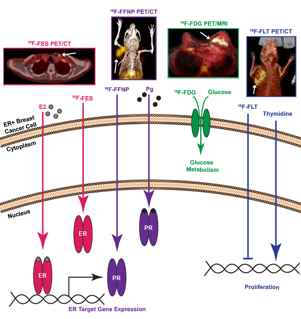
Our research is focused on advancing the use of molecular imaging to better understand the biology of breast cancer including its response to targeted drug therapies and early identification of drug resistance. Predictive and pharmacodynamic molecular imaging biomarkers may help physicians choose the best treatment tailored to each patient based on their specific tumor signaling characteristics and ultimately improve survival. Specifically, we are interested in quantitative imaging of steroid hormone receptors (estrogen and progesterone receptor) and their associated signaling pathways since the majority of breast cancer deaths occur in patients with metastatic estrogen receptor alpha (ER) positive disease.

Our experimental approaches span the translational continuum from preclinical (cell-based assays and tumor xenograft models) to clinical studies. We are currently focused on using 18F-labeled radiopharmaceuticals and multimodality imaging with positron emission tomography (PET) and computed tomography (CT) or magnetic resonance imaging (MRI). Additionally, optical imaging (fluorescence and bioluminescence) is an important tool used for our preclinical work.

My research program focuses on molecular imaging biomarkers of the estrogen signaling pathway in estrogen receptor (ER) positive breast cancer. Positron emission tomography (PET) imaging using a radiolabeled estrogen, 18F-fluoroestradiol (FES), is a read-out of the ligand binding functionality of ER. An axial fused PET/computed tomography (CT) image demonstrates FES uptake in a left supraclavicular lymph node in a patient with metastatic ER+ breast cancer (pink, arrows 1, 2, from left). Progesterone receptor (PR) is a classic estrogen-regulated ER target gene. PET imaging with a radiolabeled progestin analog, 18F-fluorofuranylnorprogesterone (FFNP) is an endogenous read-out of ER functional transcriptional activity. A maximum intensity projection fused PET/CT image demonstrates FFNP uptake in a PR+ mammary carcinoma cell line grown as a murine xenograft located in the right thoracic mammary fat pad (purple, arrows 3,4 from left). Estrogen signaling also increases glucose uptake through genomic and non-genomic mechanisms. PET imaging with a radiolabeled glucose analog, 18F-fluorodeoxyglucose (FDG), reflects tumor glucose metabolism in vivo. An axial fused PET/magnetic resonance imaging (MRI) image demonstrates avid FDG uptake in a patient with inflammatory left breast cancer and abnormal left internal mammary lymph node (green, merging arrows). Estrogen signaling culminates in increased tumor cell proliferation which can be measured by PET imaging with a radiolabeled thymidine analog, 18F-fluorothymidine (FLT). A maximum intensity projection fused PET/CT image demonstrates FLT uptake in a tumor xenograft located in the right thoracic mammary fat pad (blue, right 2 arrows).

Contact Information


Department of Radiology
University of Wisconsin School of Medicine and Public Health
600 Highland Avenue
Madison, WI  53792-3252