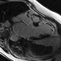
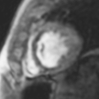
Cardiac Magnetic Resonance (CMR) Imaging
- ischemic heart disease
- viability assessment
- stress testing
- evaluating cardiac function and determining causes of heart failure
- assessing congenital heart disease
- characterizing cardiac tumors
- confirming causes of non-ischemic cardiomyopathy
- dilated cardiomyopathy
- hypertrophic cardiomyopathy
- arrhythmogenic right ventricular cardiomyopathy/dysplasia
- cardiac amyloidosis
- cardiac sarcoidosis
- left ventricular non-compaction cardiomyopathy
- iron-overload cardiomyopathy
- myocarditis
- endomyocarial fibrosis
- evaluating pericardial diseases including constrictive pericarditis
- assessing the pulmonary veins prior to pulmonary vein isolation in patients with atrial fibrillation
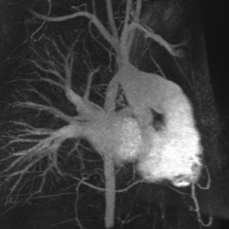
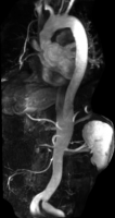
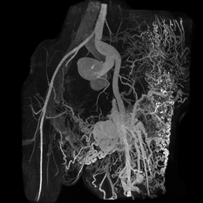
Magnetic Resonance Angiography (MRA)
Chest, Abdomen and Pelvis
- pulmonary embolus
- aortic aneurysms
- dissections, intramural hematoma and penetrating atherosclerotic ulcers
- vasculitis
- renal artery stenosis, including fibromuscular dysplasia
- mesenteric ischemia
- pelvic congestion syndrome
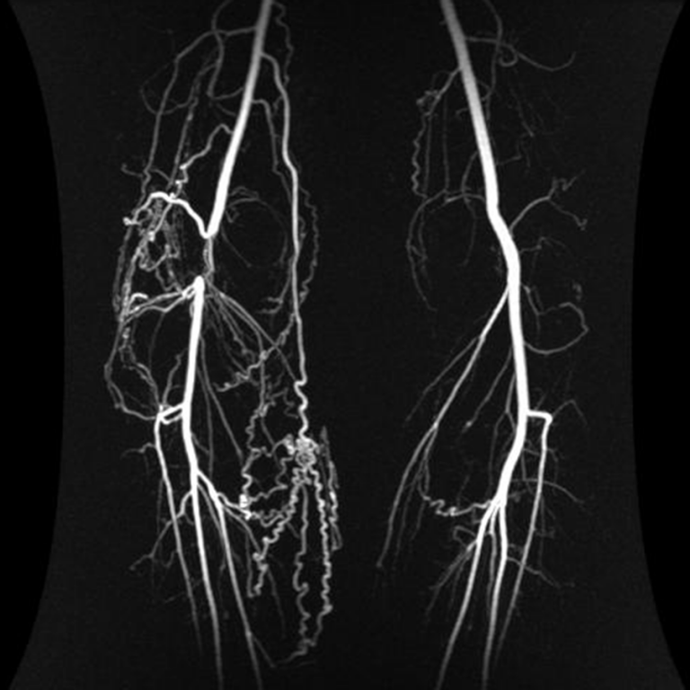
Extremities
- acute and chronic peripheral vascular disease
- vascular malformations
- popliteal entrapment syndrome
- thoracic outlet syndrome
- deep venous thrombosis, including May-Thurner syndrome and Paget-Schroetter Syndrome
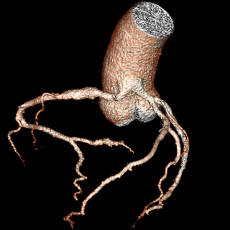
Coronary and Cardiac Computed Tomography Angiography (CCTA)
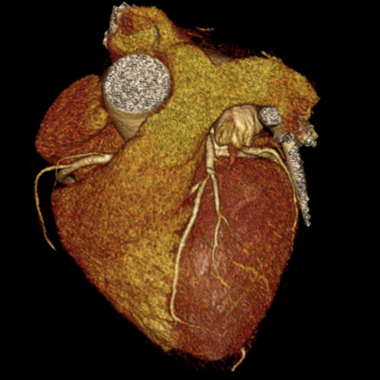
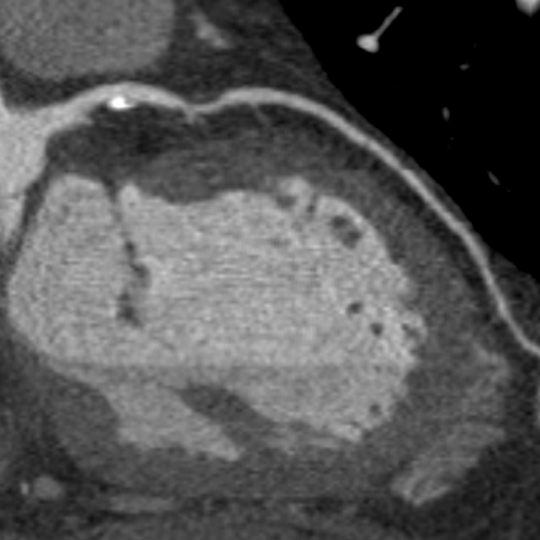
- coronary artery disease
- anomalous coronary arteries
- congenital heart disease
- evaluation of patients prior to transcatheter aortic valve replacement (TAVR)
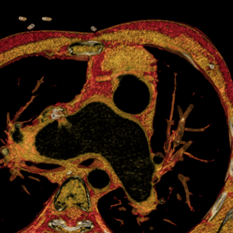
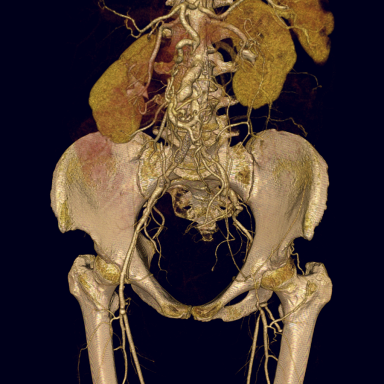
Computed Tomography Angiography (CTA)
Chest, Abdomen and Pelvis
- pulmonary embolus
- aortic aneurysms, prior to and following open or endovascular repair
- dissections, intramural hematoma, and penetrating atherosclerotic ulcers
- vasculitis
- renal artery stenosis, including fibromuscular dysplasia
- mesenteric ischemia
Extremities
- acute and chronic peripheral vascular disease
- vascular malformations
- thoracic outlet syndrome

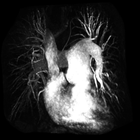
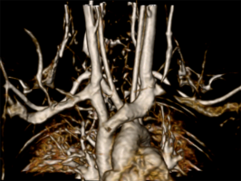
3D Lab
The dedicated team of cardiovascular imaging post-processors provide advanced 3D visualization and post-processing for optimal visualization of anatomy, pathology and surgical planning. This includes visualization and quantification of four-dimensional (4D) flow MRI and generating patient-specific 3D printed models to assist with complex surgery.