Cover Article
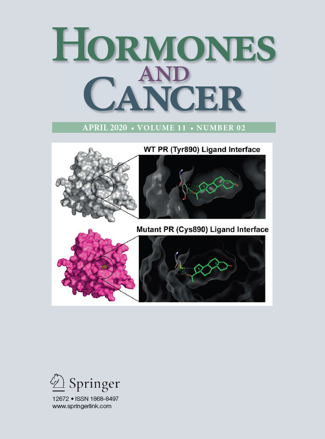
The article below was featured on the cover of the April 2020 issue of Hormones and Cancer.
 Fowler AM; Salem K; DeGrave M; Ong IM; Rassman S; Powers GL; Kumar M; Michel CJ; Mahajan AM. Progesterone receptor gene variants in metastatic estrogen receptor positive breast cancer. Hormones and Cancer. 2020 Jan; 11(2):63-75.
Brief Summary: Tumor mutations in the gene encoding estrogen receptor alpha (ESR1) have been identified in metastatic breast cancer patients with endocrine therapy resistance. However, relatively little is known about the occurrence of mutations in the progesterone receptor (PGR) gene in this population. The study objective was to determine the frequency and prognostic significance of tumor PGR mutations for patients with estrogen receptor (ER)-positive metastatic breast cancer. Thirty-five women with metastatic or locally recurrent ER+ breast cancer were included in this IRB-approved, retrospective study. Targeted next-generation sequencing of the PGR gene was performed on isolated tumor DNA. Associations between mutation status and clinicopathologic factors were analyzed as well as overall survival (OS) from time of metastatic diagnosis. The effect of the PGR variant Y890C (c.2669A>G) identified in this cohort on PR transactivation function was tested using ER−PR− (MDA-MB-231), ER+PR+ (T47D), and ER+PR− (T47D PR KO) breast cancer cell lines.
Review Articles
Keigley, Quinton J.; Fowler, Amy M.; O’Brien, Sophia R.; Dehdashti, Farrokh. Molecular Imaging of Steroid Receptors in Breast Cancer. Cancer Journal (United States). 2024 May; 30:142-152.
Brief Summary: Steroid receptors regulate gene expression for many important physiologic functions and pathologic processes. Receptors for estrogen, progesterone, and androgen have been extensively studied in breast cancer, and their expression provides prognostic information as well as targets for therapy. Noninvasive imaging utilizing positron emission tomography and radiolabeled ligands targeting these receptors can provide valuable insight into predicting treatment efficacy, staging whole-body disease burden, and identifying heterogeneity in receptor expression across different metastatic sites. This review provides an overview of steroid receptor imaging with a focus on breast cancer and radioligands for estrogen, progesterone, and androgen receptors.
Kamaraju S; Fowler AM; Tarima S; Chaudhary LN; Burkard M; Giever T; Cheng YC; Parkes A; Lange C; Pipp-Dahm M; Hegeman R; Siddiqui N; Stella A; Rajguru S; Twaroski K; Zurbriggen L; Jorns J; Rui H; Keigley QJ; Perlman SB; Salem K; Bradshaw TJ; Sahmoud T; Wisinski K. A Phase II Trial of OnapriStone and FuLvestrant for Patients with ER+ and HER2- Metastatic Breast Cancer. Clinical Breast Cancer. 2024.
Brief Summary: The SMILE study is a multi-institutional phase II clinical trial to determine the efficacy and safety of an antiprogestin, onapristone, in combination with fulvestrant as second-line therapy for patients with ER+, PgR+/-, HER2- metastatic breast cancer. This study was terminated early and herein, we report patient characteristics, and outcomes.
Salem K; Reese RM; Alarid ET; Fowler AM. Progesterone receptor mediated regulation of cellular glucose and 18F-fluorodeoxyglucose uptake in breast cancer. Journal of the Endocrine Society. 2023; 7(2):bvac186.
Brief Summary: Positron emission tomography imaging with 2-deoxy-2-[18F]-fluoro-D-glucose (FDG) is used clinically for initial staging, restaging, and assessing therapy response in breast cancer. Tumor FDG uptake in steroid hormone receptor–positive breast cancer and physiologic FDG uptake in normal breast tissue can be affected by hormonal factors such as menstrual cycle phase, menopausal status, and hormone replacement therapy. The purpose of this study was to determine the role of the progesterone receptor (PR) in regulating glucose and FDG uptake in breast cancer cells.
Note: This manuscript was chosen for the Endocrine Society Thematic Issue on Women’s Health 2023 guided by Altmetric Attention Scores and Feature Article designations.
Parent EE; Fowler AM. Nuclear receptor imaging in vivo – clinical and research advances. Journal of the Endocrine Society. 2022 Dec; 7(3):bvac197.
Brief Summary: Nuclear receptors are transcription factors that function in normal physiology and play important roles in diseases such as cancer, inflammation, and diabetes. Noninvasive imaging of nuclear receptors can be achieved using radiolabeled ligands and positron emission tomography (PET). This quantitative imaging approach can be viewed as an in vivo equivalent of the classic radioligand binding assay. A main clinical application of nuclear receptor imaging in oncology is to identify metastatic sites expressing nuclear receptors that are targets for approved drug therapies and are capable of binding ligands to improve treatment decision-making. Research applications of nuclear receptor imaging include novel synthetic ligand and drug development by quantifying target drug engagement with the receptor for optimal therapeutic drug dosing and for fundamental research into nuclear receptor function in cells and animal models. This mini-review provides an overview of PET imaging of nuclear receptors with a focus on radioligands for estrogen receptor, progesterone receptor, and androgen receptor and their use in breast and prostate cancer.
Fowler AM; Strigel RM. Clinical advances in PET/MRI for breast cancer. The Lancet Oncology. 2022; 23(1)e32-e43.
Brief Summary: Imaging is paramount for early detection of breast cancer, clinical staging, informing management decisions, and directing therapy. Positron emission tomography/magnetic resonance imaging (PET/MRI) is a quantitative hybrid imaging technology that combines metabolic, functional PET data with anatomic detail and functional perfusion information from MRI. The clinical applications for which PET/MRI may be beneficial for breast cancer is an active area of research. This Review discusses the rationale for using PET/MRI for patients with breast cancer and summarizes the clinical evidence across the spectrum of diagnosis, staging, prognosis, tumor phenotyping, and treatment response assessment. Continued development and approval of targeted radiopharmaceuticals, together with radiomics and automated analysis tools, will further expand the opportunity for PET/MRI to provide added value for breast cancer imaging and patient care.
Fowler AM; Kumar M; Henze Bancroft L; Salem K; Johnson JM; Karow J; Perlman SB; Bradshaw TJ; Hurley SA; McMillan AB; Strigel RM; Measuring glucose uptake in primary invasive breast cancer using simultaneous time-of-flight breast PET/MRI: a method comparison study with prone PET/CT. Radiology Imaging Cancer. 2021; 3(1):e200091.
Brief Summary: To compare the measurement of glucose uptake in primary invasive breast cancer using simultaneous, time-of-flight breast PET/MRI with prone time-of-flight PET/CT.
Note: This manuscript was highlighted by a commentary article:
Mankoff DA; Surti S. PET/MRI for Primary Breast Cancer: A Match Made Better by PET Quantification? Radiology Imaging Cancer. 2021;3(1):e200150.
Kumar M; Salem K; Michel CJ; Jeffery JJ; Yan Y; Fowler AM. 18F-Fluoroestradiol positron emission tomography imaging of activating estrogen receptor alpha mutations in breast cancer. The Journal of Nuclear Medicine. 2019; 60:1247-1252.
Brief Summary: The purpose of this study was to determine the effect of estrogen receptor-α gene (ESR1) mutations at the tyrosine (Y) 537 amino acid residue within the ligand binding domain on 18F-fluoroestradiol (18F-FES) binding and in vivo tumor uptake compared with wild-type (WT)-estrogen receptor α (ER).
Salem K; Kumar M; Yan Y; Jeffery JJ; Kloepping KC; Michel CJ; Powers GL; Mahajan AM; Fowler AM. Sensitivity and isoform specificity of 18F-fluorofuranylnorprogesterone for measuring progesterone receptor protein response to estradiol challenge in breast cancer. The Journal of Nuclear Medicine. 2019; 60(2):220-226.
Brief Summary: The purpose of this study was to evaluate the ability of 21-18F-fluoro-16α,17α-[(R)-(1′-α-furylmethylidene)dioxy]-19-norpregn-4-ene-3,20-dione (18F-FFNP) to measure alterations in progesterone receptor (PR) protein level and isoform expression in response to 17β-estradiol (E2) challenge.
Note: This manuscript was selected as the Featured Basic Science Article for this issue and was highlighted by an invited perspective:
Paquette M, Turcotte E. Measuring estrogen receptor functionality using progesterone receptor PET imaging: rising to the (estradiol) challenge! The Journal of Nuclear Medicine. 2019; 60(2):218–219.