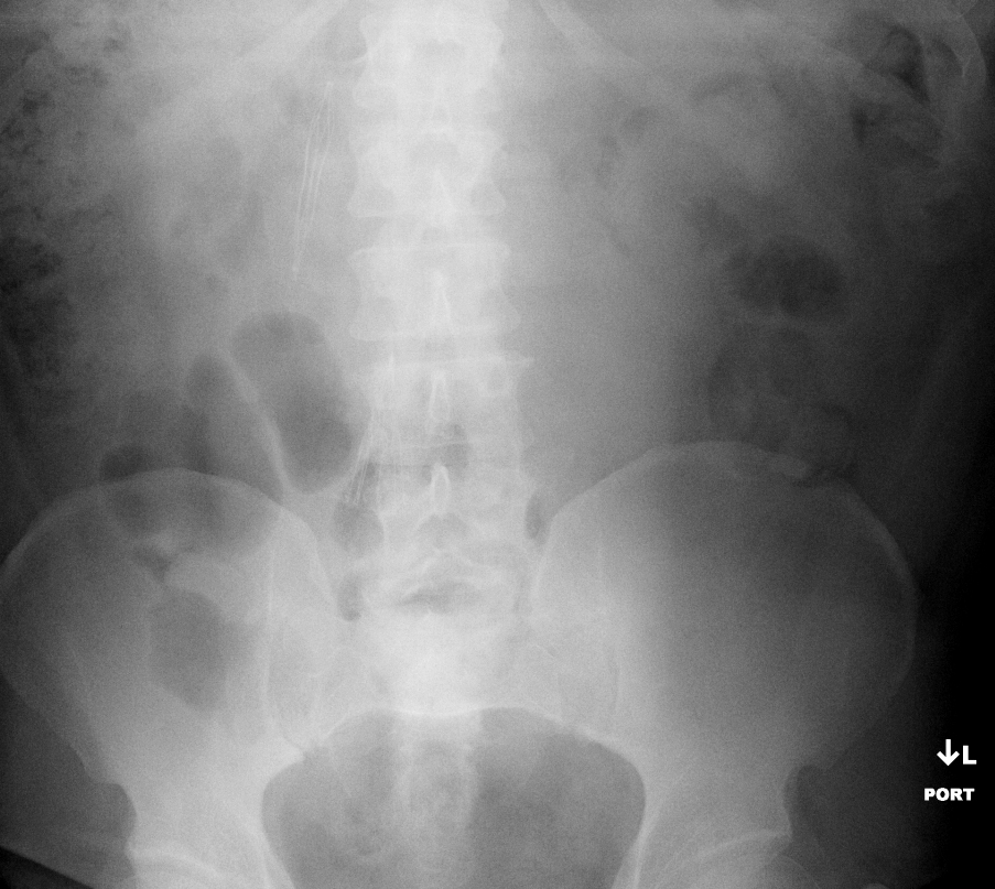
History: 43 yo male with history of lymphoma and recurrent pulmonary embolism.
Solution: This abdominal film identifies two misplaced IVC filters. The inferior filter was positioned in the right common iliac vein. Thus, the patient had recurrent PE from the left venous system, which spurred the clinicians to request placement of a second filter. The second filter did not fully deploy because it was placed in the right gonadal vein and thus did not have room to expand. Please note that neither of these filters were placed by radiology. This patient did ultimately get a third IVC filter that was positioned in the appropriate location. The renal veins typically empty into the IVC at the L1/L2 level and therefore, that is typically about where the top of a correctly deployed filter should be. In this case, both filters are obviously out of position and this case shows why a venocavogram or other assessment of the venous anatomy should be performed prior to filter placement.
