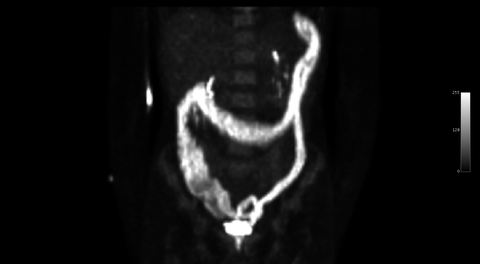
History: 55 yo male with colon cancer. Staging PET.
Solution: Ulcerative colitis.
This PET/FDG scan was performed for colon cancer staging in a middle ages male with a history of ulcerative colitis. There was a colonoscopy performed 4 days earlier that showed inflammation throughout the colon, a normal terminal ileum, and a small colon cancer. The finding of diffuse colonic uptake of this degree is certainly unusual and reflects the active inflammation associated with the ulcerative colitis. UC is associated with a 3-5% risk of developing colonic adenocarcinoma, but the most common cause of death in these patients is actually toxic megacolon.
Differentiating between US and Crohns can be challenging, but typically UC starts at the rectum and progresses proximally, while Crohns starts at the IC valve and is associated with skip lesions from mouth to anus. Both have a double peak of incidence in the young (20-40) and older (60-70).
