Treatment of nonionic radiographic contrast material extravasation

Revised 6/9/2025

Background

Contrast extravasation, also known as contrast infiltration, refers to the unintentional leakage of intravenously injected contrast media from a vein into the surrounding soft tissues. It is a rare phenomenon happening in 0.1 – 1.2% (1/1000 – 1/83) of all CT contrast injections. Extravasations with gadolinium-based contrast media are much less common than those seen after injection with iodinated contrast media but can happen.

Most contrast extravasations resolve without complications. Severe extravasation events are extremely rare happening in <<1% of all extravasations. Severe extravasations can induce compartment syndrome, a painful condition caused by a build-up of pressure in a closed muscle or tissue compartment, potentially restricting blood flow and leading to tissue necrosis, and nerve damage. Other severe reaction sequelae include skin blistering and necrosis. While it was initially hypothesized that larger volumes of extravasated contrast were more likely to cause compartment syndrome, there have been studies showing that small volumes can also cause it, particularly when it happens in less capacious volumes of tissues, such as the wrist or foot. Low- and iso-osmolar contrast agents (e.g., iohexol and iodixanol, respectively) have an even lower chance of causing a severe extravasation event as compared to older high-osmolar agents (e.g., diatrizoate) which are no longer used intravenously.

Extravasations are more common in patients who cannot communicate effectively (e.g., infants, patients with altered consciousness), are severely ill or debilitated, have altered circulation in the injected extremity (e.g., lymphedema, phlebitis, acute trauma), had radiation to an injected extremity, or are injected in the hand, foot, or ankle.

Power injected contrast via flow-rate approved central lines and PICCs also vastly decreases the incidence of extravasations.

Workflow

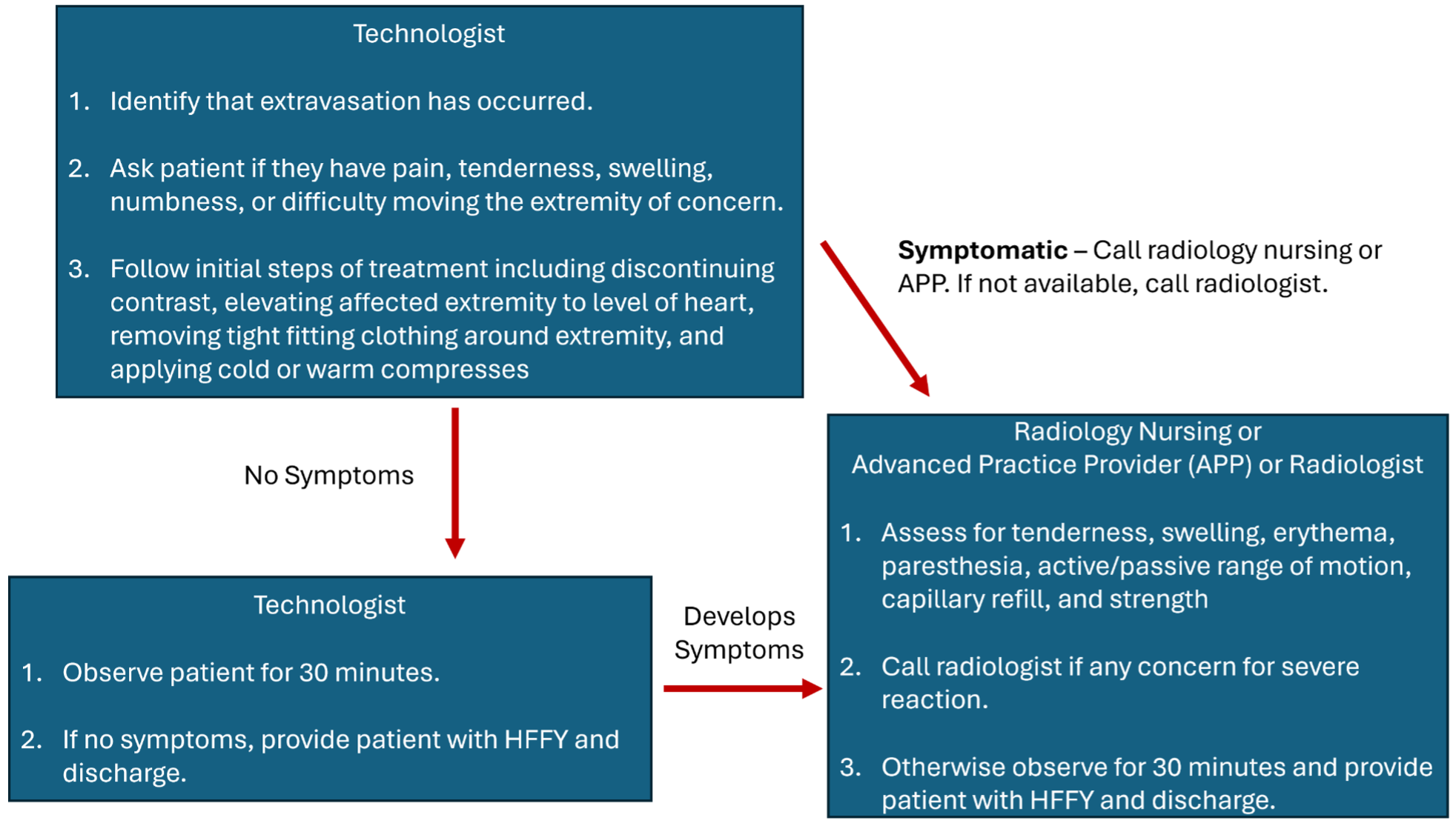
In most patients, initial swelling and tenderness resolve within hours to days after extravasation. Most patients recover without any clinically significant sequelae. If a contrast extravasation happens, use these guidelines (regardless of contrast agent):

Initial Identification:

An imaging technologist is the first health care provider likely to recognize a contrast extravasation.

  1. Technologist should immediately stop the contrast injection.
  2. Technologist gathers the following information:
    • Site of extravasation
    • Contrast agent involved
    • Approximate volume of extravasated contrast.
  3. Technologist should ask the patient the following questions:
    • Do you have any pain in the injected extremity? If yes, what is your pain level on a scale of 1-10 with 10 being the worst pain you’ve ever experienced?
    • Do you have and numbness in the injected extremity?
    • Does the injected extremity feel weaker?

Technologist should begin the following initial treatments for all extravasations:

  1. Immediately discontinue the contrast injection when a problem is detected.
  2. Place patient in an observation area.
  3. Elevate effected extremity to or above the level of the heart.
  4. Remove any tight-fitting clothing above the injection site.
  5. If patient prefers, give patient a hot or cold compress for the affected site, although this is not specifically indicated.

If patient does not indicate any pain or discomfort on initial assessment: Technologists observe patient for at least 30 minutes. If patient does not have worsening symptoms, patient can be discharged using the discharge protocol below.

If patient appears acutely symptomatic on initial survey or patient develops worsening symptoms after 5 minutes of observation/treatment or the technologist feels unsure of their assessment (e.g., patient mental status altered and unable to convey symptoms): Technologist should call a radiology nurse or advanced practice practitioner to assess the patient. The technologist should readily convey any clinical information they have gathered once additional health-care providers arrive. If a nurse or APP is also concerned for compartment syndrome, the radiologist should be notified to assess the patient.

If a nurse or APP is unavailable, a radiologist (attending or trainee) should assess the patient.

We stress that compartment syndrome almost always takes time to develop, on the order of hours to days.

Nurse/APP/Radiologist Assessment:

  • Perform a physical exam to assess capillary refill, muscle strength, numbness, skin findings (e.g., edema, ulceration).
  • If nurse or APP is concerned for compartment syndrome or skin blistering, call radiologist to assess patient.
  • Plastic Surgery Consultation: If a radiologist is concerned for compartment syndrome, immediate plastic surgery consultation should be performed for any of the following scenarios:
    • Severe pain or progressive swelling and or pain
    • Skin ulceration, blistering, or necrosis
    • Altered tissue perfusion as evidenced by decreased capillary refill distal to the injection site
    • Change in sensation in the affected limb
    • Worsening passive or active range of motion
  • Surgical consultation based on volume of extravasated contrast is not recommended since most patient with large volume extravasations do not develop severe complications, even when distal to the elbow. Therefore, surgical consultation should be based on signs and symptoms rather than an absolute volume threshold.

Discharge Protocol:

  • For Outpatients
    • Technologists should give patient the Contrast Extravasation Health Facts for You (HFFY).
    • Health care providers involved in patient’s care should verbally stress that if patients’ symptoms worsen, they should immediately contact a healthcare provider as indicated in the HFFY.
  • For Inpatients
    • Technologist should place the Contrast Extravasation Order Set (NURCOM0092) which communicates to inpatient teams on further guidance.
  • Technologist should place a HERO (PSN) for each extravasation event.
  • For cases in which radiologists, nurses, or APPs are involved, a short note should be documented in the patient’s medical record by the respective provider.

Contrast Extravasation Workflow Summary

Bicarb Protocol

Interventional Radiology Bicarb Protocol for CIN

Indication

Contrast induced or associated AKI is felt to be a real albeit rare entity.  The most important risk factor seems to be pre-existing severe renal insufficiency (AKI or eGFR<30mL/min/1.73m2.  The major preventative action to mitigate the risk of CIN is to provide intravenous volume expansion (see Hydration protocol).  One possible protocol would be 0.9% saline at 100 ml/hr, beginning 6-12 hrs before contrast and continuing for 4-12 hours after contrast in the inpatient setting.

A study from JAMA printed in May of 2004 indicates that hydration with a bicarbonate solution may better prevent contrast induced nephropathy than NS hydration. Most renal failure is associated with metabolic acidosis and low urinary pH. NS may contribute to acidosis while the bicarbonate solution will buffer the pH.3.  Subsequent studies have challenged this practice, and results are not definitive at this time.  However, given the low cost and lack of side effects, it is unlikely to be harmful and could be added to the hydration regimen.  Other practices, such as administration of N-acetyl cysteine, are not supported by the literature.  Cessation of nephrotoxic medications may also be helpful when feasible.

Administration/Dosing

Solution: 150 mEq NaHCO3 in 1000cc D5W

1 hour prior to contrast: Initial IV bolus – 3ml/kg/hour x 1 hour

After 1 hour bolus: 1ml/kg/hour during contrast exposure and 6 hours post contrast.

References

  1. Levy EM, Viscoli CM, Hurwitz RI. The effect of acute renal failure on mortality: a cohort analysis. JAMA. 1996; 275: 1489-1494.
  2. McCullough PA, Wolyn R, Rocher LL, et al. Acute renal failure after coronary intervention. Am J Med.1997; 103: 368-375.
  3. Merten GJ, et al. Prevention of contrast-induced nephropathy with sodium bicarbonate: a randomized controlled trial. JAMA. 2004; 291: 2328-2334.
  4. 2004/Lisa Semmann, R.N., M.S., Interventional Radiology NP

Prophylaxis Policy for Patients Allergic to IV Contrast

7/2020

What patients require premedication prior to IV contrast?

A prior allergic-like or unknown type reaction to the same class of contrast medium is considered the greatest risk factor for predicting future adverse events. Premedication prior to administration of intravenous contrast is recommended in this group of patients.

In general, patients with unrelated allergies are at a 2- to 3-fold increased risk of an allergic-like contrast reaction, but due to the modest increased risk, restricting contrast medium use or premedicating solely on the basis of unrelated allergies is not recommended. Patients with shellfish or povidone-iodine (e.g., Betadine®) allergies are at no greater risk from iodinated contrast medium than are patients with other allergies (i.e., neither is a significant risk factor).

In patients with reported prior contrast allergy or reaction:

  1. Ascertain the specific details of the prior reaction.  Determine:
    1. If the patient had a true reaction to intravascular contrast material
    2. Type of contrast used (if available)
    3. Type of reaction and severity
    4. Treatment and its result
    5. Level of patient anxiety
  2. In addition to intravascular contrast administration, the following scenarios should be considered in patients with contrast allergy:
    1. Administration of oral contrast.  1-2% of oral contrast could be absorbed.  So for someone who has a severe contrast allergy, they should get dilute barium as an oral contrast agent.  If prior mild reaction and minimum risk of perforation/leak, no need to premedicate.  For moderate reactions or risk of peritoneal leak, consider premedication or dilute barium alternate oral contrast.
    2. Arthrograms.  Because allergic-like reactions are dose-independent, and arthrograms use a needle and cross the bloodstream, use the same premedication strategy for arthrograms that is used for intravenous media. For patients with known contrast allergies, appropriate to premedicate or switch to US if possible.
    3. Hysterosalpingograms: In patients with patent fallopian tubes, there is peritoneal spill of contrast.  Intraperitoneal contrast is absorbed (intraperitoneal=intravascular).  Because of the dose independence, premedication for HSGs should be performed in patients with known contrast allergies.
  3. Determine if an examination without contrast can provide adequate diagnostic information.
  4. If still convinced of the need for intravascular contrast, the service requesting  the scan should:
    1. Contact the radiologist scheduled on service for the proposed date of the study (GI/GU, CT, Angiography, Neuroradiology, or on-call resident for overnight/weekend Emergency Department patients).  If the staffing schedule is not available for the proposed date of the examination, then the Chief of the GI/GU Service (Dr. Perry Pickhardt), CT Service (Dr. Meghan Lubner), Angiography/Interventional Service (Dr. Orhan Ozkan), or Neuroradiology Service (consultant for the day) should be contracted.
    2. Discuss why an alternative imaging method will not suffice
    3. If the risk is determined to be acceptable; schedule the contrast examination and transmit prophylaxis instructions.
  5. Routine Contrast Reaction Prophylaxis (12 hour):
    1. Methylprednisolone (Medrol)
      1.  32 mg by mouth at 12 and 2 hours before contrast injection.
    2. Diphenhydramine (Benadryl):
      1. 50 mg intramuscular or oral 1 hour before contrast injection OR
      2. 50 mg (or 25 mg per height/weight indication) intravenously 15-20 min before contrast injection
        Note:  The Benadryl must be used with the proviso that the patient does not drive a car or operate heavy machinery 4-6 hours after the Benadryl is administered.
        NEJM 1987; 317: 845-849.
    3. If a patient is unable to take oral medication, consider the 5 hr regimen detailed in #6.  Alternatively the methylprednisolone may be substituted with 200 mg hydrocortisone IV at 13, 7, and 1 hour before contrast administration. If a patient is allergic to diphenhydramine in a situation where diphenhydramine would otherwise be considered, an alternate anti-histamine without cross-reactivity may be considered, or the anti-histamine portion of the regimen may be dropped.
  6. Rapid Contrast Reaction Prophylaxis (5 hours):
    1. To be used only in a truly urgent situation after discussion between the requesting service and the appropriate radiology service.
    2. The requesting service must place a note in HealthLink outlining the necessity of contrast-enhanced examination and the rapid prohpylaxis protocol.
    3. Hydrocortisone:
      1. 200 mg intravenously 5 and 1 hour before contrast injection
    4. Diphenhydramine (Benadryl):
      1. 50 mg intravenously 1 hour before contrast injection
  7. In addition to steroid premedication, changing the culprit iodinated agent if known may decrease the risk of repeat allergic reaction. Consider substitution of an iso-osmolar agent (iodixanol) if a low osmolar agent was previously administered.

Greenberger PA et al. Emergency administration of radiocontrast media in high-risk patients. J Allergy CLin Immunol. 1986; 77(4): 630-634.

Mervak BM, Cohan RH, Ellis JH, Khalatbari S, Davenport MS.  Premedication administered 5 hours before CT compared with a Traditional 13-hour Oral regimen. Radiology 2017; 285(2), 425-433..

McDonald JS, Larson NB, Kolbe AB et al. Prevention of Allergic-like Reactions at Repeat CT: Steroid Pretreatment versus Contrast Material Substitution. Radiology 2021; 000:1-8.

Metformin-Containing Medications

List of diabetic medications known to contain Metformin

  • ActoPlus Met®
  • ActoPlus Met XR®
  • Avandamet®
  • Fortamet®
  • Glucophage®
  • Glucophage XR®
  • Glucovance®
  • Glumetza®
  • Invokamet®
  • Janumet®
    Janumet XR®
  • Jentadueto®
  • Kazano®
  • Kombiglyze XR®
  • Metaglip®
  • PrandiMet®
  • Riomet®
  • Xigduo XR

Who must have a creatinine level prior to IV contrast?

3/2022

Reference: ACR Manual on Contrast Media, 2021

All inpatients require a current (within one week) creatinine level or estimated glomerular filtration rate (eGFR) prior to an IV contrast-enhanced CT.

Outpatients and ED patients being scheduled for a CT with IV contrast will not require a serum creatinine unless they meet one of the following criteria:

  • Personal history of renal disease, including:
    • Known chronic kidney disease (CKD)
    • Remote history of AKI
    • Dialysis
    • Kidney surgery or kidney transplant
    • Kidney ablation
    • Albuminuria
  • History of diabetes mellitus (optional)
  • Metformin or metformin-containing drug combinations

These parameters are intended to serve as guidelines. Obviously, medical necessity may mandate administering contrast without a current measure of renal function.