Background
Contrast extravasation, also known as contrast infiltration, refers to the unintentional leakage of intravenously injected contrast media from a vein into the surrounding soft tissues. It is a rare phenomenon happening in 0.1 – 1.2% (1/1000 – 1/83) of all CT contrast injections. Extravasations with gadolinium-based contrast media are much less common than those seen after injection with iodinated contrast media but can happen.
Most contrast extravasations resolve without complications. Severe extravasation events are extremely rare happening in <<1% of all extravasations. Severe extravasations can induce compartment syndrome, a painful condition caused by a build-up of pressure in a closed muscle or tissue compartment, potentially restricting blood flow and leading to tissue necrosis, and nerve damage. Other severe reaction sequelae include skin blistering and necrosis. While it was initially hypothesized that larger volumes of extravasated contrast were more likely to cause compartment syndrome, there have been studies showing that small volumes can also cause it, particularly when it happens in less capacious volumes of tissues, such as the wrist or foot. Low- and iso-osmolar contrast agents (e.g., iohexol and iodixanol, respectively) have an even lower chance of causing a severe extravasation event as compared to older high-osmolar agents (e.g., diatrizoate) which are no longer used intravenously.
Extravasations are more common in patients who cannot communicate effectively (e.g., infants, patients with altered consciousness), are severely ill or debilitated, have altered circulation in the injected extremity (e.g., lymphedema, phlebitis, acute trauma), had radiation to an injected extremity, or are injected in the hand, foot, or ankle.
Power injected contrast via flow-rate approved central lines and PICCs also vastly decreases the incidence of extravasations.
Workflow
In most patients, initial swelling and tenderness resolve within hours to days after extravasation. Most patients recover without any clinically significant sequelae. If a contrast extravasation happens, use these guidelines (regardless of contrast agent):
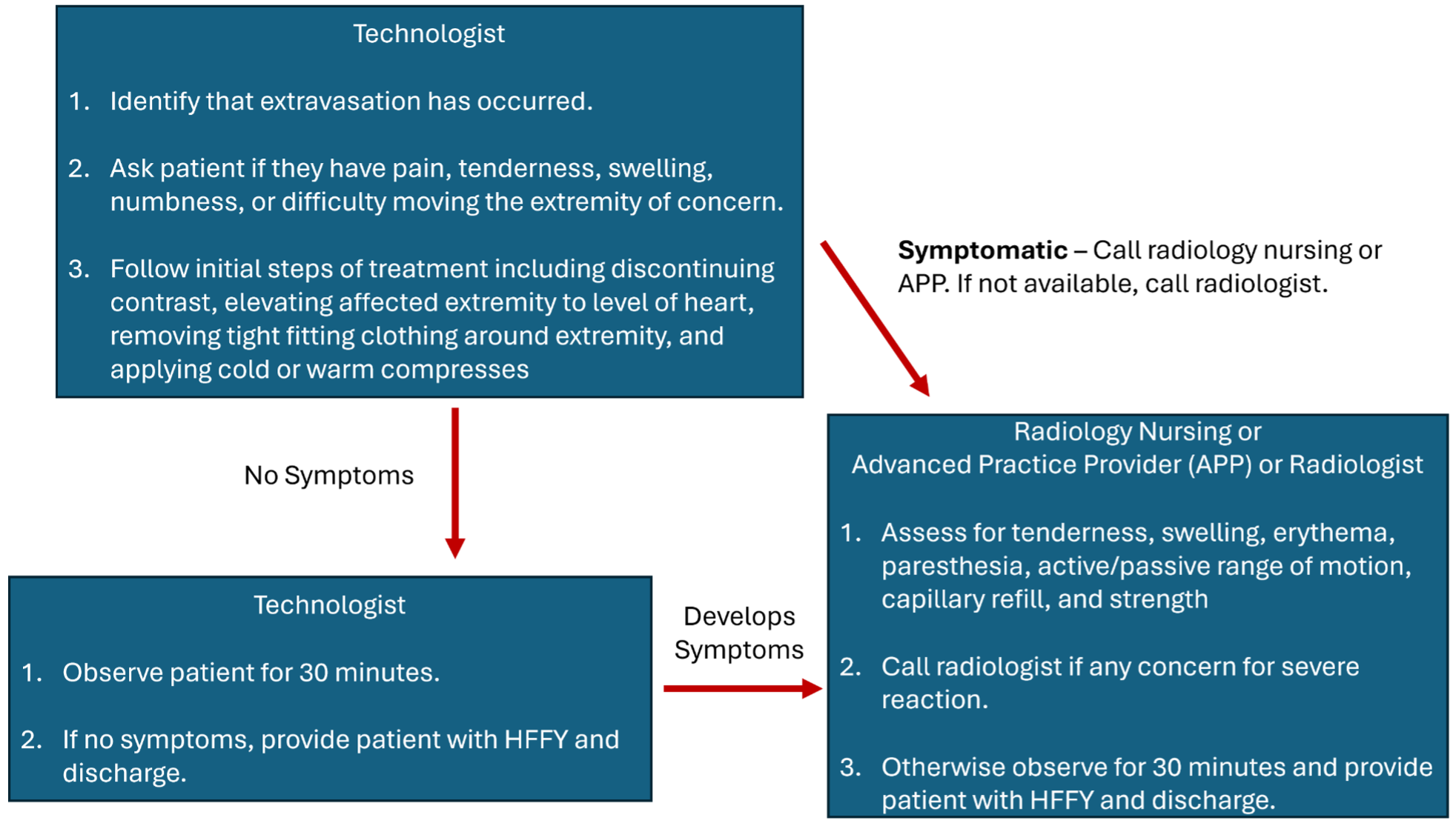
Initial Identification:
An imaging technologist is the first health care provider likely to recognize a contrast extravasation.
- Technologist should immediately stop the contrast injection.
- Technologist gathers the following information:
- Site of extravasation
- Contrast agent involved
- Approximate volume of extravasated contrast.
- Technologist should ask the patient the following questions:
- Do you have any pain in the injected extremity? If yes, what is your pain level on a scale of 1-10 with 10 being the worst pain you’ve ever experienced?
- Do you have and numbness in the injected extremity?
- Does the injected extremity feel weaker?
Technologist should begin the following initial treatments for all extravasations:
- Immediately discontinue the contrast injection when a problem is detected.
- Place patient in an observation area.
- Elevate effected extremity to or above the level of the heart.
- Remove any tight-fitting clothing above the injection site.
- If patient prefers, give patient a hot or cold compress for the affected site, although this is not specifically indicated.
If patient does not indicate any pain or discomfort on initial assessment: Technologists observe patient for at least 30 minutes. If patient does not have worsening symptoms, patient can be discharged using the discharge protocol below.
If patient appears acutely symptomatic on initial survey or patient develops worsening symptoms after 5 minutes of observation/treatment or the technologist feels unsure of their assessment (e.g., patient mental status altered and unable to convey symptoms): Technologist should call a radiology nurse or advanced practice practitioner to assess the patient. The technologist should readily convey any clinical information they have gathered once additional health-care providers arrive. If a nurse or APP is also concerned for compartment syndrome, the radiologist should be notified to assess the patient.
If a nurse or APP is unavailable, a radiologist (attending or trainee) should assess the patient.
We stress that compartment syndrome almost always takes time to develop, on the order of hours to days.
Nurse/APP/Radiologist Assessment:
- Perform a physical exam to assess capillary refill, muscle strength, numbness, skin findings (e.g., edema, ulceration).
- If nurse or APP is concerned for compartment syndrome or skin blistering, call radiologist to assess patient.
- Plastic Surgery Consultation: If a radiologist is concerned for compartment syndrome, immediate plastic surgery consultation should be performed for any of the following scenarios:
- Severe pain or progressive swelling and or pain
- Skin ulceration, blistering, or necrosis
- Altered tissue perfusion as evidenced by decreased capillary refill distal to the injection site
- Change in sensation in the affected limb
- Worsening passive or active range of motion
- Surgical consultation based on volume of extravasated contrast is not recommended since most patient with large volume extravasations do not develop severe complications, even when distal to the elbow. Therefore, surgical consultation should be based on signs and symptoms rather than an absolute volume threshold.
Discharge Protocol:
- For Outpatients
- Technologists should give patient the Contrast Extravasation Health Facts for You (HFFY).
- Health care providers involved in patient’s care should verbally stress that if patients’ symptoms worsen, they should immediately contact a healthcare provider as indicated in the HFFY.
- For Inpatients
- Technologist should place the Contrast Extravasation Order Set (NURCOM0092) which communicates to inpatient teams on further guidance.
- Technologist should place a HERO (PSN) for each extravasation event.
- For cases in which radiologists, nurses, or APPs are involved, a short note should be documented in the patient’s medical record by the respective provider.
Contrast Extravasation Workflow Summary