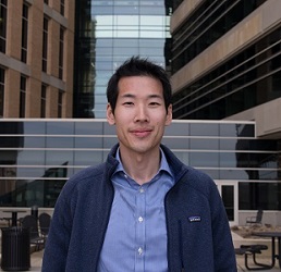
Christian Park, a fourth year medical student working with Dr. Alan McMillan, has been selected for a Radiological Society of North America (RSNA) Medical Student Research Grant. The grant is for the project “Ultra Low Dose PET/MRI Imaging of Crohn’s Disease using a novel Deep Learning Reconstruction Method.”
RSNA has established this grant to, “increase the opportunities for medical students to have a research experience in medical imaging and to encourage them to consider academic radiology as an important option for their future.”
According to Park, reduction of ionizing radiation exposure is important for any patient, but is a critical factor for Crohn’s Disease (CD) patients. Typically, CD patients are quite young at diagnosis, will begin receiving radiology treatment early, and may require monitoring throughout their lives. There is currently no cure for CD. Magnetic Resonance Imaging (MRI) is an excellent imaging modality, with superior contrast compared to Computed Tomography (CT) and does not expose patients to ionizing radiation. However, it can be difficult to discern between active disease and fibrotic tissue using MR alone. A different imaging procedure, Simultaneous Positron Emission Tomography (PET), can highlight physiologically active tissue, allowing detection of inflammatory lesions, though does deliver a higher amount of radiation exposure. This research project focuses on using state of the art Deep Learning algorithms to reduce the dose of PET tracer required, so that the ionizing radiation exposure of a simultaneous PET/MR approaches that of an abdominal plain film.
By sparing the radiation required for one examination, more studies can be performed without surpassing the radiation exposure from previous methods, allowing for closer monitoring of disease activity. Conversely, more frequent scans may indicate remission achievement, thereby allowing the reduction or cessation of drug intervention, which can also have significant side effects and substantial financial cost. Prompt detection of inflammation, which can occur in the absence of clinical symptoms such as pain or nausea, may facilitate rapid treatment and support prevention of serious complications. Therefore, with the development of new low dose imaging techniques, imaging techniques that were previously reserved for diagnosis or extent of disease evaluation can now be used as screening examinations, providing valuable information to better guide and inform future care.
In radiology, Artificial Intelligence (AI), and more specifically Deep Learning, has been of significant interest, as evidenced by the rapid growth of research and incredible amount of investment by corporations and institutions. AI adaptations for a multitude of different applications across radiology are being developed, from computer aided diagnosis (CAD) to improving PACS to EMR crosstalk. Ultimately, continued AI research will accelerate progress in the field of radiology and result in improved diagnosis and treatment for patients. In this project, AI is being used to reduce radiation exposure, by training an algorithm to recognize the features of a full-dose image from an acquired low-dose image thereby allowing a low-dose image to be used in place of the conventional full-dose images.
“It is an honor for me to receive this grant from the RSNA so that I have the opportunity to pursue my research interests in Artificial Intelligence with Dr. McMillan at the University of Wisconsin,” said Park. “Having recently matched into an academic radiology residency program, I hope to continue my research throughout my career and contribute to the field of radiology. AI has enormous potential and only through continued progress and efforts can we discover all the ways in which we can improve the lives of patients.”