Every so often, an idea comes along that has the potential to radically change the status quo. For digital subtraction angiography, that time has come. The always impressive team of Charles Mistretta, Ph.D., of the Imaging Sciences Section, and Charles Strother, M.D., emeritus professor in the Neuroradiology Section, just received a perfect score from the National Institutes of Health (NIH) evaluators and a $1.2 million grant to continue their pioneering research in 4D digital subtraction angiography and 4D fluoroscopy. NIH review committees rarely award a perfect score to even the best grant proposal, and this reflects NIH’s enthusiastic support for Dr. Strother and Dr. Mistretta’s research.
Digital subtraction angiography (DSA) is a type of fluoroscopy technique used in interventional radiology to visualize blood vessels in a bony or dense soft tissue environment. Fluoroscopy is a technique that can allow physicians to obtain real-time, moving images of internal structures of a patient. The simplest form of fluoroscopy images are X-rays. DSA images are produced using contrast medium by subtracting a pre-contrast image from later images once the contrast medium has been introduced into the structure. Often, the medium of choice for visualizing blood vessels is iodine.
This grant is the direct result of a long-time partnership between Drs. Mistretta and Strother. Their research hopes to improve the safety and diagnostic capabilities of DSA. The pair began working on DSA techniques shortly after Dr. Strother came to UW-Madison in 1976, and made great strides in improving current techniques. Upon his return to UW-Madison in 2006, DSA techniques had evolved into 3D vascular reconstructions, and Dr. Strother had ideas about how to further enhance the impact DSA could have on the imaging world. After consulting with Dr. Mistretta, the concept of 4D DSA and 4D fluoroscopy was born.
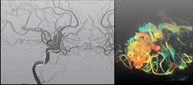
4D DSA is an X-ray technique that adds an additional dimension to traditional DSA by producing a time series of 3D image volumes at high frame rates. Using this technique, the examination time is greatly reduced, which exposes patients to a far lower dose of radiation. This particular grant from NIH is specifically funding the validation of the diagnostic and therapeutic capabilities of 4D DSA and 4D fluoroscopy, which will advance this technique toward implementation on a broad scale.
This incredible ground-breaking study has the potential to change DSA imaging throughout the field of radiology. We are anxiously awaiting more exciting news from this project.