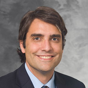
 Innovation in medical imaging often takes years of behind-the-scenes hard work and effort. Diego Hernando, PhD and Scott Reeder MD, PhD have been researching an innovative method of measuring liver iron concentration for over a decade, and their findings are ready to be shared with the world in their paper, “Multicenter Reproducibility of Liver Iron Quantification with 1.5-T and 3.0-T MRI,” which was recently published in Radiology.
The origin of this study begins in 2010 when Dr. Hernando was hired as an Assistant Scientist in the Department of Radiology’s Imaging Sciences section. He began working with Dr. Reeder, who was already researching diffuse liver disease. They were both curious if there was a correlation between the measurement of liver iron concentration and R2*, the rate of decay of the MRI signal.
Dr. Hernando said, “In the scientific community, there was a lot of doubt about the reproducibility of using R2*. Some people thought since R2* depends on background magnetic field homogeneity, presence of fat or noise, or so on, that R2* is an inherently poorly reproducible measurement.” However, Drs. Reeder and Hernando believed it was important to measure liver fat and R2* jointly because one without the other would lead to biased results. They first completed a lot of technical development. Dr. Reeder said, “We measure the MRI signal using complex numbers, meaning we use both the magnitude and phase of the signal, and when these complex signals are used to measure R2*, that measurement is intrinsically insensitive to errors due to noise. Older R2* methods ignore the signal phase, which leads to bias, or an error, in the measurement of R2* and therefore estimates of liver iron concentration.”
In 2012, Drs. Hernando and Reeder performed a single-center study, funded by a $100,000 two-year grant from WARF, to evaluate their hypothesis that if you corrected R2* for all the relevant factors, you could use different acquisition parameters and still produce the same results. They recruited 50 subjects, testing different acquisition protocols. “Testing different protocols is really important in the context of quantitative imaging methods, because a lot of times, you can’t always keep your acquisition parameters fixed, because of hardware and software differences, vendor differences, etc.,” said Dr. Hernando. From their single-center study at the University of Wisconsin – Madison, they were able to prove their hypothesis using a single vendor, GE, at 1.5-T and 3.0-T.
From there, they gained the confidence to pursue a multicenter study. For this expansion, they kept their focus on patients with iron overload but expanded their research to other centers with different MRI vendors and different patient populations. They joined forces with the University of Texas–Southwestern which uses Philips MRI systems, Johns Hopkins University which uses Siemens, and Stanford University, which uses GE, and has more pediatric patients with iron overload. “This multi-center study has successfully demonstrated the viability of R2*-based iron quantification in the liver as a reproducible biomarker across vendors, protocols, and between 1.5T and 3.0T. This now means that a patient could be scanned on almost any platform, and we can compare results between different scanners and different sites anywhere in the world,” said Dr. Reeder.
Not only is this project a testament to clinical innovation in the measure of liver iron overload, but it is also a testament to collaborative research in the Department of Radiology. This decade-long project started at the beginning of Dr. Hernando’s career at UW-Madison when he was an Assistant Scientist until now when he is an Associate Professor with his very own lab, Quantitative Imaging Methods Lab. Dr. Reeder remarked, “It’s been an absolute pleasure to see this brilliant young scientist blossom into an internationally renowned scientist and engineer. Diego has extraordinary technical capabilities and an amazing knack for working with clinicians to understand the current clinical limitations and apply technical solutions with advanced imaging technologies.”
Congratulations to Drs. Reeder and Hernando for their publication in Radiology! We cannot wait to see how this research translates to the clinical space!