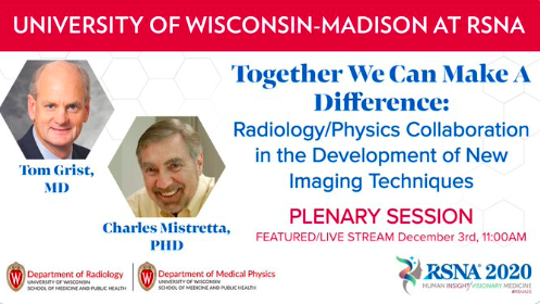
 As part of RSNA 2020, Professor and Radiology Department Chair Thomas Grist, MD, FACR, and Professor Charles Mistretta, PhD, gave a collaborative lecture. The lecture, titled “Together We Can Make a Difference: Radiology/Physics Collaboration in the Development of New Imaging Techniques” focused on X-ray, MRI and CT techniques for diagnostic and interventional angiographic applications, as well as the history that led to these developments. Angiography is a medical imaging technique used to visualize the lumen of blood vessels, particularly arteries, veins, and heart chambers.
RSNA 2020 is a virtual event this year, being held from Sunday, November 29 through Saturday, December 5. This year’s focus is human insight and visionary medicine. The meeting provides an opportunity for radiology professionals to enhance their skills; connect with peers; and see the latest research, education, and technology.
Dr. Grist’s research focuses on developing and applying advanced, noninvasive MRI techniques to diagnose and provide therapy for cardiovascular disorders. Dr. Grist is the John H. Juhl Professor of Radiology, Medical Physics, and Biomedical Engineering. His physics background, and his interprofessional collaborative research team, help him bridge the gap from lab work to clinical practice. Dr. Grist also helped establish the Imaging Sciences Center in the Wisconsin Institutes for Medical Research (WIMR), which focuses on developing imaging technologies and then translating these technologies for clinical use.
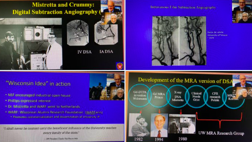 Dr. Mistretta is a Professor Emeritus in Medical Physics, Radiology, and Biomedical Engineering. His research focuses on safe diagnostic angiographic interventions and time-resolved angiography, and has resulted in accelerated imaging techniques such as TRICKS, VIPR, and HYPR. He was also part of the research group that developed digital subtraction angiography (DSA). His current research interests include non-invasive techniques for MRI’s of the cardiovascular system, quantitation of flow, coronary artery imaging and flow measurement, and MR myocardial perfusion.