Last fall Giuseppe Toia, MD, an expert on CT imaging and the medical director of the UW/GE Healthcare CT Protocol Collaboration served as a panelist during the American Journal of Roentgenology (AJR) forum on Photon-Counting Detector CT (PCD-CT).
PCD-CT directly converts x-rays to electronic signal, allowing for substantial gains in spatial resolutions, contrast, noise reduction, and multienergy reconstruction. Previous CT technology uses energy-integrating detectors (EID-CT), following a two-step process of converting x-rays to light, then converting light to electronic signal.
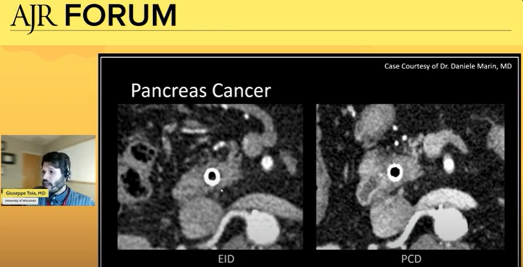
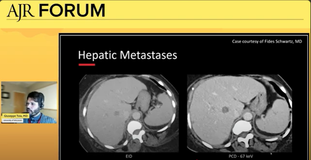
During the panel, Dr. Toia illustrated clinical applications of PCD-CT for abdominal imaging. He highlighted its exciting potential to better monitor pancreas cancer and detect metastases in organs like the liver. He also shared that it’s beneficial in imaging the overall anatomy of the pancreas.
|
|
|
The panelists discussed possible challenges with adopting PCD-CT into clinical practice. One concern is efficiency; with both increased spatial resolution content and quantity of images to review radiologists. Dr. Toia emphasized that protocols must be specially designed to balance efficiency with work throughput.
While these concerns are valid, PCD-CT holds promising potential to improve patient treatment and outcomes in clinical settings. Dr. Toia looks forward to a more widespread use of the technology, citing “It’s clear by everyone’s presentation today that this technology leverages and shows us things that we have not seen before. Not just ‘we can see something better and be more comfortable about calling it’ but clearly that we’re seeing pathology we’ve never seen before.”
Watch the entire AJR Forum featuring Dr. Toia and co-panelists Timothy Amrhein, MD; Fernando Kay, MD, PhD; and Fides Schwartz, MD to learn more about Photon-Counting Detector CT.