Presenter: Timothy Szczykutowicz, Ph.D.
An educational exhibit examining techniques to reduce dosage in several CT modalities won a cum laude award at RSNA ’14, one of four UW presentations to earn the accolade. The group was also invited to present their findings to RadioGraphics, the official RSNA journal. Assistant Professor Timothy Szczykutowicz, Ph.D. presented the exhibit, and additional UW authors include Assistant Professor Tabby Kennedy, M.D., and former Neuroradiology Fellow Sara Nace, M.D.
Szczykutowicz began by illustrating the major difference between MDCT and CACT. “MDCT images are acquired using a ‘donut’ geometry, where the gantry spins 360 degrees around the subject,” said Szczykutowicz. “CACT is acquired on a C-Arm, where the gantry only goes roughly 220 degrees and travels back and forth.” While CACT scans irradiate the patient slightly less than MDCT scans by virtue of a smaller scanning angle, that’s just the tip of the iceberg.
The true difference-maker is the VOI mode, a special modality only available in CACT. “Most radiologists don’t know about this, because it’s so new,” remarked Szczykutowicz. For example, in traditional CT imaging, to image the heart, a radiologist needs to radiate the entire cross section of the patient’s chest. Sending x-rays only at the heart would result in a truncated projection, an incomplete reconstruction of the image.
However, improvements in reconstruction algorithms within this VOI mode can eliminate some of those artifacts, allowing a radiologist to target a sub-volume within a patient’s anatomy. “Why is that advantageous? If I just cared about your heart, why would I want to radiate the entire slice through your chest? The end result is a smaller [radiation] dose to the patient,” said Szczykutowicz.
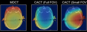
The group had absolute dose data to back up their assertions, taken from high-contrast temporal bone imaging, pictured above. A patient being scanned using VOI mode receives one-sixth the amount of radiation compared to undergoing a scan using MDCT, according to Szczykutowicz. That remarkable reduction has lead Siemens and other manufacturers to explore implementation of VOI mode on CACT, a measure that could bring VOI to hospitals and clinics nationwide.