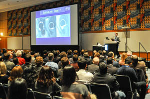
 A standing-room-only crowd was eager to hear Donna Blankenbaker, M.D., speak on one of her favorite subjects: hip imaging. Dr. Blankenbaker began with an overview of the labrum, noting that the high density of nerve endings causes labral tears to be quite painful. While anterior-to-anterior tears are common, posterior tears are much rarer, and tend to happen in young athletes. Magnetic resonance (MR) arthrography is currently the preferred technique for detecting labral tears, however, there are several situations that make diagnosis difficult.
One challenge is the chondrolabral junction. While it can appear to be a tear on MR, physicians must be careful to avoid over-diagnosis. Using all imaging planes available can help radiologists make the correct decision, according to Dr. Blankenbaker. Another frequent misstep comes in diagnosing the perilabral recess as a tear, when it is in fact a natural structure. The ligamentum teres, or round ligament, can also present problems. Since it is a small structure, it can be hard to determine a tear via imaging, although Dr. Blankenbaker noted that putting the leg in traction can make for much easier diagnosis.
Another problematic feature of the hip joint is the possible presence of hip plicae. A plica is a fold of membrane most commonly found in the knee. Plicae are present in about 50% of the population and are thought to be the remnants of embryonic connective tissue that failed to fully resorb during fetal development. Hip plicae are commonly seen in MR arthrography, and may cause pain, so they are occasionally excised.
Join the conversation on Twitter @UWiscRadiology, #RSNA15