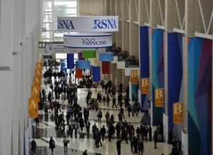
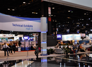
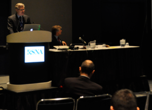
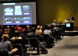
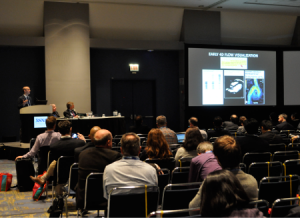
UW Radiology continued its success at the annual meeting of the Radiologic Society of North America (RSNA), the largest medical conference in the world. The 2015 edition of the conference was marked by nearly 50 awards for UW posters or presentations, including magna cum laude awards for four separate presentations. Faculty such as Fred T. Lee Jr, M.D., and Ken Schreibman, Ph.D./M.D., gave marquee talks on some of their favorite topics: ablation and the musculoskeletal system, respectively. Even several residents and fellows joined in, contributing to award-winning papers and posters.
Special congratulations are in order for Meghan Lubner, M.D., and Perry Pickhardt, M.D., who each garnered six or more awards over the week-long conference. Other top performers include Scott Sheehan, M.D., and Tabassum Kennedy, M.D., who each received multiple awards for papers or presentations.
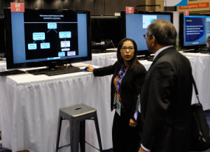
UW Radiology’s CT protocol development was a prime topic of discussion at GE’s RSNA booth, as over 200 sites in the US are now using UW-developed protocols, in addition to dozens of international sites. Additionally, Kaiser Permanente, an Oakland-based health care system, has committed to use UW CT protocols in a pilot program at 10 of their hospitals. This may result in a nationwide standardization of their CT protocols based on the work performed at UW.
Presentation Summaries
- Fred T. Lee Jr Touts the Benefits of Microwave AblationUW is a nationwide leader in microwave ablation, thanks to the efforts of the Tumor Ablation Lab. Led by Fred. T. Lee Jr, M.D., and Christopher Brace, Ph.D., the lab has produced full-fledged ablation systems, dozens of patents, and a spinoff company called Neuwave Medical. Dr. Lee shared the insights gained from years of developing and performing MW ablations, in a talk entitled “The Case for Microwave Ablation.â€
- Dr. Blankenbaker Reviews Frequent Hip MRI MisstepsA standing-room-only crowd was eager to hear Donna Blankenbaker, M.D., speak on one of her favorite subjects: hip imaging. Magnetic resonance (MR) arthrography is currently the preferred technique for detecting labral tears; however, there are several situations that make diagnosis difficult.
- Mai Elezaby Reports on Usefulness of BI-RAD SystemSince the Breast Imaging-Reporting and Data (BI-RAD) System went into effect in 2008, it has helped standardize the reporting of mammograms through a six-tier system. Mai Elezaby, M.D., conducted a retrospective study on the effectiveness and efficacy of the BI-RADS system, presented at RSNA 15.
- Elizabeth Burnside Merges Imaging and GenomicsElizabeth Burnside, M.D., is part of a multi-institutional team investigating the connection between imaging features and genetic profile, in order to predict the reoccurrence of breast cancer. The team reviewed computer extracted MRI features and gene expression tests, finding that the risk of recurrence was most strongly indicated by the size and shape of the tumor. While there was a limited data pool for the study, the team made compelling observations about how internal tumor architecture may play a biological role in reoccurrence.
- Dr. Schreibman Returns to ASRT@RSNAKen Schreibman, M.D./Ph.D., continued his award-winning lecture series with his 3rd straight ASRT@RSNA appearance, speaking this year about how to correctly image foot and ankle fractures. Ankle fractures are the 4th most common extremity fracture, and there are three main varieties, each with their own unique imaging needs. Dr. Schreibman reviewed the anatomy of the ankle and demonstrated how to position the patient to obtain the most useful images, all the while providing interesting information about the historical treatment of ankle injuries.
Awards
** Magna cum laude
* Cum laude
‡ Certificate of Merit
§ Selected for Submission to RadioGraphics
Φ Trainee Research Prize
Sarah Averill, M.D.
- ‡ Global Health: Improving Early Detection of Breast Cancer in Rural Areas. From Community Education to Ultrasound Exams, A Model for Reducing Barriers to Breast Care
- ** Multi-system Imaging Features of Cystic Fibrosis in Adults
Brian Chan, M.D.
- *§ MRI of Pediatric Bone Marrow: The Normal, the Abnormal, and the Exceptions
Zachary Clark, M.D.
- ** When Less is More: Exploiting Vessel Sparsity to improve 4D CEMRA of Dural AV Fistulas
Lori Mankowski Gettle, M.D.
- ठInfiltrative Renal Lesions in Adults РSpectrum of Disease
Kara G. Gill, M.D.
- *§ MRI of Pediatric Bone Marrow: The Normal, the Abnormal, and the Exceptions
Eric Hartman, M.D.
- *§ ‘Up Against the Wall’ Assessing Cerebrovascular Stroke Risks by Combining 4D Flow MRI and Arterial Wall Imaging
Kevin M. Johnson
- *§ ‘Up Against the Wall’ Assessing Cerebrovascular Stroke Risks by Combining 4D Flow MRI and Arterial Wall Imaging
- ** When Less is More: Exploiting Vessel Sparsity to improve 4D CEMRA of Dural AV Fistulas
Tabby Kennedy, M.D.
- ‡ Imaging Findings of Non-cutaneous Melanoma
- ** Multi-system Imaging Features of Cystic Fibrosis in Adults
David Kim, M.D.
- * Heterotopic Pancreatic Rests: Imaging Features, Complications, and Unifying Concepts
Kenneth S. Lee, M.D.
- * What’s that Sound? Sonographic Evaluation of Common Shoulder Pathologies with MRI Correlation
Meghan G. Lubner, M.D.
- * Anorectal Evaluation at CT Colonography: Pathology and Pitfalls
- ‡ Extra-Osseous Ewing Sarcoma: Spectrum of Disease With Pathologic Correlation
- * Heterotopic Pancreatic Rests: Imaging Features, Complications, and Unifying Concepts
- ‡ Imaging Findings of Non-cutaneous Melanoma
- ** Mibs and Mabs: Intra-abdominal Complications of Targeted Cancer Therapies
- ** Multi-system Imaging Features of Cystic Fibrosis in Adults
- * Oncogenic Infections: A Review
- *§ Vasculitis in the Emergency Room Setting
Matthew Lee, M.D.
- ‡§ What’s that Sound? Sonographic Evaluation of Common Shoulder Pathologies with MRI Correlation
Jie Nguyen, M.D.
- *§ MRI of Pediatric Bone Marrow: The Normal, the Abnormal, and the Exceptions
Perry J. Pickhardt, M.D.
- * Anorectal Evaluation at CT Colonography: Pathology and Pitfalls
- ‡ Extra-Osseous Ewing Sarcoma: Spectrum of Disease With Pathologic Correlation
- * Heterotopic Pancreatic Rests: Imaging Features, Complications, and Unifying Concepts
- ‡ Imaging Findings of Non-cutaneous Melanoma
- ठInfiltrative Renal Lesions in Adults РSpectrum of Disease
- ** Multi-system Imaging Features of Cystic Fibrosis in Adults
Susan Rebasemen, M.D.
- *§ MRI of Pediatric Bone Marrow: The Normal, the Abnormal, and the Exceptions
Humberto Rosas, M.D.
- ** Long Head Biceps Tendon Injuries of the Shoulder: Injury Biomechanics, and the Role of Imaging in Clinical Management
Elizabeth Sadowski, M.D.
- ‡ Fertility Preservation in Women with Gynecological Cancers: Review of Fertility Sparing Techniques and Role of MR Imaging
Andrew Scarano, M.D.
- ‡ ‘Up Against the Wall’ Assessing Cerebrovascular Stroke Risks by Combining 4D Flow MRI and Arterial Wall Imaging
Scott Sheehan, M.D.
- ** Long Head Biceps Tendon Injuries of the Shoulder: Injury Biomechanics, and the Role of Imaging in Clinical Management
- ‡ What’s that Sound? Sonographic Evaluation of Common Shoulder Pathologies with MRI Correlation
Jason W. Stephenson, M.D.
- ** Long Head Biceps Tendon Injuries of the Shoulder: Injury Biomechanics, and the Role of Imaging in Clinical Management
Patrick Turski, M.D., FACR
- ‡ ‘Up Against the Wall’ Assessing Cerebrovascular Stroke Risks by Combining 4D Flow MRI and Arterial Wall Imaging
- ** When Less is More: Exploiting Vessel Sparsity to improve 4D CEMRA of Dural AV Fistulas
Oliver Wieben, Ph.D.
- ** When Less is More: Exploiting Vessel Sparsity to improve 4D CEMRA of Dural AV Fistulas
Vincenzo Wong, M.D.
- ‡ Imaging Findings of Non-cutaneous Melanoma
Yijing Wu, Ph.D.
- *§ ‘Up Against the Wall’ Assessing Cerebrovascular Stroke Risks by Combining 4D Flow MRI and Arterial Wall Imaging
- ** When Less is More: Exploiting Vessel Sparsity to improve 4D CEMRA of Dural AV Fistulas