
Next-Gen GE Scanner Comes Online
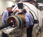
UW Radiology and GE Healthcare have a decade-old partnership, dating back to collaborations on CT protocols during the early 2000s. This relationship was strengthened by the April installation of the SIGNA PET/MR scanner, the first of its kind from GE.
While previous PET/MR combination scanners exist, none are as advanced as the SIGNA. “The combination of the updated design with new detectors makes this more sensitive than any scanner on campus,” said Alan McMillan, Ph.D. “It’s a pretty unique instrument.”
The state-of-the-art detectors are called silicon photomultipliers, replacing the photomultiplier tubes that are so common on older PET scanners. They improve both image timing and resolution, but the real coup is in their compatibility with MR. The tube technology was unsuited for the magnetic and radiofrequency fields in an MR scanner, and forced PET imaging to be used in conjunction with CT.
“CT gives you good anatomic imaging, but you don’t get a lot of contrast in soft tissue,” said McMillan. “Now with MR, it’s very sensitive with soft tissue, and we’re merging this with PET. That gives us very specific information.”
The benefits become apparent when applied to a disease like irritable bowel syndrome (IBS). IBS causes inflammation, which results in increased metabolism in affected areas. Combining the soft-tissue imaging of MR with PET molecules that seek out high-metabolic cells can give clinicians an accurate picture of the damage, at a low radiation dose.
However, the most innovative uses of PET/MR are yet to come, according to McMillan. “Increasing the sensitivity while still reducing dose might allow us to use PET/MR where it’s been impossible before.”
Burnside Group Takes Aim at “Radiogenomics”
In an age where websites customize ads to a user’s browsing history, and taste in music can be deduced through algorithms, it’s only natural that medical imaging will follow a similar path towards personalization. Elizabeth Burnside, M.D., MPH, M.S. believes patient data can be used to tailor screening methods to individual patients, using imaging biomarkers and patient history.
“We now can access electronic health records and imaging in a highly confidential manner. We can use that huge amount of data to make better healthcare decisions,” said Dr. Burnside. Mortality reduction with breast cancer screening has been well established but personalized strategies to minimize false positives and maximize accurate diagnoses are the next step.
“Most of our grants look at informatics to predict risk. We are now incorporating genetics merged with imaging features to better predict risk, called radio-genomics,” said Dr. Burnside. Her research group specializes in personalizing breast imaging strategies using electronic health data. “You take the individual characteristics and risk, the performance of the technology, and see which exam is best. For example, in the case of women with dense breast tissue, 3D mammography or ultrasound may be more effective than traditional mammography,” said Dr. Burnside.
In the future, they hope this screening philosophy can be extended to other disorders such as Alzheimer’s Disease. Using neuroimaging to identify early biomarkers for Alzheimer’s builds on the idea of establishing a screening culture similar to breast cancer’s. “If you catch it early, you can intervene and improve outcomes,” said Dr. Burnside.
The end goal for this breast cancer initiative is an application that could evaluate a patient’s history and recommend the most effective exams.
“I think this is where the imaging field is going. If we can personalize our approaches and prove we’re affecting outcomes positively and minimizing harms, then screening policies will be clearer and less controversial,” said Dr. Burnside.
Imaging Informatics Group Building Data Processing Cloud
The marriage of information technology and healthcare is unique, combining the data-heavy nature of modern healthcare and the stringent requirements of the federal Health Insurance Portability and Accountability Act of 1996 (HIPAA). This means that any data processing involving patient records must happen internally, or be anonymized before it leaves the secure hospital network.
For radiology, internal data processing can mean creating virtual machines. These machines perform a variety of functions, from 3D image processing to analysis of patient data. Creating a virtual machine has been a manual process, from provisioning storage and memory, to installing operating systems and archiving information. However, Vice Chair of Informatics Gary Wendt, M.D., MBA, believes that an internal computing cloud can solve many of these problems.
The new cloud will build virtual machines, and automatically provide storage, memory, and archive services. This automation will allow researchers to process non-anonymized data quickly and efficiently.
“You can pick from a menu, it’s almost like going to a deli and pointing at what toppings you want for a sandwich,” said Dr. Wendt. “It’s not aimed to replace a desktop, but it’s aimed to provide services, both for clinical and translational applications.”
The first phase of the project was deployed in mid-July, and is undergoing internal testing to refine the request and approval process.
Molecule Manufacturing Facility Speeds Bench to Bedside Transition

Among the myriad additions associated with WIMR II, the one with the biggest impact might be the Radiopharmaceutical Manufacturing Facility (RPF). This state-of-the-art laboratory can manufacture molecular imaging agents to an extremely high level of precision, opening up a world of possibilities for UW scientists.
“The FDA requirements for making these new molecular agents have changed over the years, and you can’t buy the agents—you have to make them yourself,” said Scott Perlman, M.D, M.S. “With this new facility, we are able to make agents that we can use internally, as well as ship throughout the state through the Wisconsin Oncology Network for Imaging eXellence (WONIX).”
The opening of the RPF dovetails nicely with the ongoing PET research at UW, and the move to WIMR II has shortened the bench-to-bedside transition. UW Radiology researchers are now physically closer to their colleagues in the Department of Medical Physics, allowing for more efficient collaboration on projects.
In addition, synthesizing molecular imaging agents on-site has the potential to drastically streamline the process of clinical trials. “You get your tracer from the RPF, you run it through quality control to ensure it’s safe, and then you can go ahead and inject it in research subjects across the hall,” said Dr. Perlman. “These are novel agents other institutions don’t have access to.”
One of the most exiting imaging agents to come out of WIMR II is a new prostate cancer PET imaging agent called DCPFPyl. Steve Y. Cho, M.D, helped to develop the drug at Johns Hopkins University and will help further develop it for clinical applications at UW. The agent targets the areas with the most aggressive disease, making it clear to clinicians where treatment is needed most. “He’ll make that agent in the RPF, and then use it on patients suffering from prostate cancer, something he wasn’t able to do before the opening of the facility,” said Dr. Perlman.