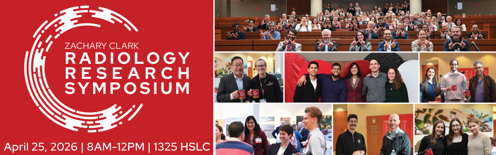
Zachary Clark Radiology Research Symposium

Program

Program Schedule

8:00 – 8:30: Continental Breakfast

8:30 – 8:45: Welcome & Introduction

8:45 – 10:15: Oral Presentations

10:15 – 10:45: Electronic Exhibits Viewing

10:45 – 11:45: Keynote Address: Dania Daye, MD, PhD on “From Innovation to Operation: Leading Through High Value Imaging”

11:45 – 12:00: Recognitions & Concluding Remarks

Keynote Speaker

Dania Daye, MD, PhD

Director, Center for High Value Imaging
Vice Chair, Practice Transformation
Associate Professor, UW Department of Radiology

“From Innovation to Operation: Leading Through High Value Imaging”

Read More in the Announcement Article

Oral Presentations

Brody Brisk, MD

Level: Fellow
Faculty Mentor: John Symanski, MD
Title: Cervical Facet Joints: Pain Improvement with Intra-articular Versus Extra-articular Injections

This is an accordion element with a series of buttons that open and close related content panels.

Abstract for Cervical Facet Joints

Citation:
Brisk BW, Ross AB, Rosas HG, Symanski JS. Cervical facet joints: pain improvement with intra-articular versus extra-articular injections. Annual SSR Meeting, Oral Presentation, March 30-April 2, 2025

Purpose:
Evaluate differences in patient pain relief after intra-articular versus extra-articular cervical facet joint corticosteroid injections.

Methods:
An electronic health record search (10/30/2016-7/17/2024) identified consecutive cervical facet joint injections performed in the radiology department. Retrospective chart review and image analysis included patient demographics, imaging guidance modality, needle approach, injected level(s), intra- versus extra-articular injection, baseline and 14-day pain score (0-10). Pre-procedure MRI characteristics including osteoarthritis severity, bone marrow edema, joint effusion/synovitis and peri-facet edema were recorded. Statistical analysis included descriptive statistics and analysis of variance to assess factors associated with pain relief.

Results:
Of 117 cervical facet joint injections, 72 with completed pain diaries were included. Single injections were performed in 39/72 (54.2%) of cases, two-level unilateral injections in 20/72 (27.8%) of cases and same level bilateral injections in 13/72 (18.1%) of cases. Fluoroscopic guidance was used in all but one case. The joint line was targeted laterally in 57/72 (79.2%) of cases, and the inferior recess targeted posteriorly in the remainder of cases 15/72 (20.8%). Intra-articular injection with a single injection or both two-site injections was achieved in 46/72 (64.9%) of cases, extra-articular injection was performed in a single injection or both two-site injections in 18/72 (25%) of cases, and in the remainder of cases 8/72 (11.1%) one injection was intra-articular and one was extra-articular. The average pain reduction at 14-days was 2.5 (SD 3.3) for intra-articular and 1.6 (SD 2.1) for extra-articular injections, though this was not statistically significant (p = 0.11). All other variables showed no association with pain relief, including needle approach, number of injections, arthritis severity, and pre-procedure MRI findings.

Conclusion:
Intra-articular injections may offer slightly better pain reduction than extra-articular injections for the cervical facet joints.

Allison Couillard, MD

Level: Resident
Faculty Mentor: Fred Lee, MD
Title: Does Histotripsy of Central Liver Tumors Cause Biliary Ductal Injuries? A Study in a Live Porcine Survival Model and Early Human Clinical Experience

This is an accordion element with a series of buttons that open and close related content panels.

Abstract for Does Histotripsy of Central Liver Tumors Cause Biliary Ductal Injuries?

Citation:
Couillard AB, Laeseke P, Knavel-Koepsel E, Ziemlewicz T, Swietlik J, Hinshaw JL, Lee FT Jr. Does histotripsy of central liver tumors cause biliary ductal injuries? A study in a live porcine survival model and early human clinical experience. SIR Annual Meeting, March 29-April 2, 2025.

Purpose:
Although rare, current locoregional therapy for hepatic lesions is associated with biliary complications such as ischemic cholangitis, radiation cholecystitis, or biloma formation. Histotripsy creates precise tissue destruction within tissues with evidence of tissue selectively, specifically sparing heavily collagenous structures, which may be beneficial when treating near critical structures such as bile ducts. The purpose of this study is to evaluate for biliary complications after histotripsy of the central liver in both porcine models and clinical patients.

Material and Methods:
Retrospective review of previously treated central liver in a non-tumored porcine survival model (n=18) was performed to assess for presence of biliary complication such as biliary dilatation, biloma, or leak immediately post-treatment and with 30-day (+/- 5 day) follow-up. For clinical patients, retrospective review of centrally treated liver lesions treated patients at a single institution was performed (n=3). Size of treatment zone, distance from common bile duct, and evidence of biliary complication was evaluated.

Results:
The average size of ablation zone in the porcine model immediately post treatment was 3.2 x 3.3 x 1.9 cm (average prescribed zone size of 2.5 cm x 2.5 cm x 2.0 cm, p > 0.05). The average distance from the central common bile duct was 8 mm +/- 2 mm. There was evidence of mild biliary dilatation in two animals (11%) at one month follow-up imaging. There was no evidence of biloma, biliary leak, hepatic abscess or gallbladder pathology. For the three clinically treated patients, the average treatment zone size was 3.2 x 2.8. 3.5 cm and 3 mm +/- 2 mm, from the common bile duct. At one month follow-up, there was no evidence of biliary complication.

Conclusion:
Histotripsy of the liver near the central bile ducts does not appear to cause significant biliary complication possibly related to the relatively tissue selective proprieties of the treatment modality.

Max Golden

Level: Medical Student
Faculty Mentor: Perry Pickhardt, MD
Title: AI-Enabled Body Composition Biomarkers at Post-Mortem CT for Enriching Autopsy Evaluation

This is an accordion element with a series of buttons that open and close related content panels.

Abstract for AI-Enabled Body Composition Biomarkers at Post-Mortem CT for Enriching Autopsy Evaluation

Citation:
Golden MV, Lee MH, Garrett JW, Berry S, Appel N, Edgar H, Pickhardt PJ. AI-Enabled Body Composition Biomarkers at Post-Mortem CT for Enriching Autopsy Evaluation. American Roentgen Ray Society; San Diego, CA, April 27-May 1, 2025.

Purpose of Study:
To correlate fully automated post-mortem CT (PMCT)-based measures of aortic calcification, skeletal muscle, and intra-abdominal fat of decedents with causes of death and comorbidities.

Materials, Methods, and Procedures:
Retrospective study of the New Mexico Decedent Image Database (NMDID) with non-contrast PMCT scans between 2010-2017. An automated pipeline of AI-driven algorithms for quantifying skeletal muscle, subcutaneous fat, visceral fat, and aortic calcification (Agatston score) from the abdominal component of PMCT scans was used. Scans with more than minimal decomposition were excluded. Cause of death was categorized as “acute” or “chronic.” CT-based “biological age” was derived using a predetermined model.

Results:
6638 decedents (mean age, 50 ± 18 [SD]; 74% male) comprised the final cohort. 80% of deaths were classified as “acute,” 10% as “chronic,” and 10% were “uncertain.” Muscle density (HU) and area at the L3 lumbar level were higher in the “acute” versus “chronic” group (26 HU vs. 18 HU, p<0.001; 192 vs. 183 cm2, p<0.001). Similarly, both muscle density and area at the L3 level were higher among those without cancer (25 HU vs. 16 HU, p<0.001; 190 cm2 vs. 169 cm2, p<0.01). Aortic Agatston scores were higher in those who died of heart disease (5120 vs. 2098, p < 0.001). Patients with diabetes had higher L3 visceral fat area (227 cm2 vs. 175 cm2, p<0.001) and lower muscle density (17 HU vs. 25 HU, p<0.001). Decedents in the chronic group had a significantly higher biological-chronological age gap compared with the acute group (median age gap, 19 years vs. 10 years; p < 0.001).

Significance of the Conclusions:
Fully automated quantitative CT-based tissue biomarkers from PMCT scans match expectations based on previous studies on living patients and correlate with acuity of death and chronic co-morbidities. These data may prove useful for enhancing autopsies, including easily deriving organ volumes and estimated weights, as well as corroborating cause of death in uncertain cases.

Abigail Johnson

Level: Medical Student
Faculty Mentor: Amy Fowler, MD, PhD
Title: Academic Clinical Practice Audit of Imaging for Pediatric Patients Presenting with Breast Symptoms

This is an accordion element with a series of buttons that open and close related content panels.

Abstract for Academic Clinical Practice Audit of Imaging for Pediatric Patients Presenting with Breast Symptoms

Citation:
Johnson AS, Chapman T, Elezaby M, Fowler AM. (2024). Academic Clinical Practice Audit of Imaging for Pediatric Patients Presenting with Breast Symptoms. [Unpublished manuscript].

Full Abstract:
Appropriate imaging evaluation and management of pediatric patients with breast symptoms is important to avoid recommending unnecessary imaging follow-up or invasive procedures that cause stress for patients and their caregivers and potential damage to the developing breast. Current practice parameters focus on adults, and there is no evidence-based consensus statement from professional societies to guide management specifically for pediatric breast masses. The objective of this study was to determine the diagnostic performance and outcomes of breast imaging in our pediatric patient population and further contribute to evidence guiding management for this clinical scenario.

This IRB-approved retrospective study was performed at a large academic medical institution with both adult and pediatric subspecialty imaging, where patients 10 years or older with breast symptoms are evaluated by breast imaging radiologists. An imaging database search was performed to identify patients less than 18 years old who underwent imaging for breast symptoms between 2013 to 2023. Patients were included in the study if initial imaging evaluation was performed at our institution during the study date range. Imaging BI-RADS assessment categories, initial management, follow-up, and final diagnoses were abstracted from the electronic medical record system. Final outcomes were based on clinical resolution of initial symptoms and signs, completion of imaging or clinical follow-up (up to 24 months), or tissue diagnosis (imaging-guided or excisional biopsy).

A total of 417 patients (340 females, 77 males; mean age 14 years old; age range 0 to 17 years) met inclusion criteria. Two patients had a personal history of non-breast cancer (medulloblastoma and leukemia, respectively), and 114 patients had a family history of cancer. The most common imaging indications were palpable lump (n=340) and pain (n=143). Ultrasound (n=425) was the primary imaging modality. Initial imaging assessments were 24% (101/429) BI-RADS 1, 24% (101/429) BI-RADS 2, 19% (81/429) BI-RADS 3, and 8.6% (37/429) BI-RADS 4A. 21 cases were assigned BI-RADS 4A on follow-up imaging. The most frequent pathology from core needle biopsy (25/37) or surgical excision (15/27) was fibroadenoma. In 323 cases with clinical/imaging follow-up or tissue diagnosis, there were no malignancies.

Appropriate management is most often clinical or imaging follow-up with infrequent indication for biopsy recommendation as the likelihood of malignancy in the pediatric population is very low. Consideration of BI-RADS 3 assessment may be an appropriate alternative to biopsy in most pediatric patients.

Adam Miller, MD

Level: Fellow
Faculty Mentor: Kenneth Lee, MD, MBA
Title: Impact of Sciatic Nerve Involvement on Return-to-Sport Time in Collegiate Football Players with Acute Hamstring Injuries: Insights from MRI

This is an accordion element with a series of buttons that open and close related content panels.

Abstract for Impact of Sciatic Nerve Involvement on Return-to-Sport Time in Collegiate Football Players with Acute Hamstring Injuries

Citation:
Miller AM, Mosiman SJ, Joachim MR, Heiderscheit BC, Lee KS. Impact of Sciatic Nerve Involvement on Return-to-Sport Time in Collegiate Football Players with Acute Hamstring Injuries: Insights from MRI. Annual SSR Meeting, Podium Presentation, March 30-April 2, 2025.

Purpose:
To assess whether the British Athletics Muscle Injury Classification (BAMIC) grade and sciatic nerve involvement on MRI at time of hamstring injury (HSI) correlate with delayed return to sport (RTS) in NCAA Division I football players.

Materials and Methods:
IRB approval and informed consent were obtained. Data was collected as part of a larger prospective cohort investigation of collegiate football players who sustained HSI. MRIs were assessed by a musculoskeletal radiologist using the BAMIC grading system. Sciatic nerve involvement was characterized by extent of adjacent edema on MRI as no contact, partial (contacted by edema) or complete (surrounded by edema). Spearman Rank correlations, ANOVA and chi-square analyses were used to determine associations between imaging characteristics, HSI severity and days missed before RTS.

Results:
74 unique HSI were included in the study. The most common primary muscle injured was the biceps femoris long head (70%) followed by the semimembranosus (18%). Intratendinous tear was the most common site of HSI (61%). BAMIC 3c was the most common HSI grade (44%) followed by BAMIC 2c (17%). Increasing BAMIC grade was associated with longer time to RTS (r = 0.42, p < 0.001). Sciatic nerve involvement was observed in 45 cases (71.4%; 20 partial
[31.7%], 25 complete [39.7%]). Complete nerve involvement (35.8 ± 15.4 days, p = 0.01) was associated with significantly longer time to RTS compared to no involvement (24.5 +/- 14.0 days). Partial involvement (30.0 ± 14.1
days, p = 0.25) did not result in significantly longer RTS. There was also a statistically greater likelihood of sciatic nerve involvement as BAMIC grade increased (p = 0.002).

Conclusion:
Increasing radiologic severity, as indicated by BAMIC grade and sciatic nerve involvement, is correlated with longer RTS intervals in HSI. Sciatic nerve involvement on MRI should be factored into the assessment of acute hamstring injuries.

References:
1.) Pollock N, James SLJ, Lee JC, Chakraverty R. British athletics muscle injury classification: a new grading system. Br J Sports Med 2014;48: 1347–1351.
2.) Day MA, Karlsson LH, Herzog MM, et al. Correlation of Player and Imaging Characteristics With Severity and Missed Time in National Football League Professional Athletes With Hamstring Strain Injury: A Retrospective Review. The American Journal of Sports Medicine. 2024;52(11):2709-2717.

Hassan Rizvi

Level: Medical Student
Faculty Mentor: Perry Pickhardt, MD
Title: Novel Abdominal CT-based Model for Defining Metabolic Syndrome Using AI-enabled Automated Biomarkers: Comparison with Clinical Criteria for Predicting Cardiovascular Event

This is an accordion element with a series of buttons that open and close related content panels.

Abstract for Novel Abdominal CT-based Model for Defining Metabolic Syndrome Using AI-enabled Automated Biomarkers

Citation:
Rizvi HA, Garrett JW, Warner JD, Kattan MW, Zea R, Liu D, Lee MH, Summers RM, Pickhardt PJ. Novel abdominal CT-based model for defining metabolic syndrome using AI-enabled automated biomarkers: comparison with clinical criteria for predicting cardiovascular events. SAR Annual Meeting. February 16-21, 2025.

Purpose:
The clinical criteria for metabolic syndrome include insulin resistance, obesity, dyslipidemia, and hypertension. However, abdominal circumference and BMI fail to capture the distribution of visceral and subcutaneous fat or account for ectopic fat depots such as hepatic steatosis and myosteatosis. Using only CT-based body composition biomarkers, we derived an imaging model for metabolic syndrome to stratify risk for major cardiovascular (CV) events and compared its performance to the clinical definition of metabolic syndrome.

Methods:
The harmonized clinical definition for metabolic syndrome required at least 3 of the following 5 criteria: A1C ≥6.5 mg/dL, T2DM, or oral hypoglycemic use; BMI ≥30 kg/m²; triglycerides ≥150 mg/dL or medication use; HDL-C <40 mg/dL for men or <50 mg/dL for women; and systolic BP ≥130 mmHg or diastolic BP ≥85 mmHg. Adults aged 18-59 years with sufficient clinical data to assess metabolic syndrome and an available abdominal CT were included. An automated pipeline of AI-enabled CT algorithms quantified skeletal muscle, abdominal fat, aortic calcification, liver, and kidneys. Multivariable survival analysis was modeled using Cox proportional hazards regression; CT biomarker selection was based on the index of prediction accuracy (IPA). The primary outcome measures were major adverse CV events (MI, CVA, CHF)

Results:
From a final cohort of 51,515 adults, 23,076 (44.8%) met the clinical definition for metabolic syndrome, while 28,439 (55.2%) did not. A total of 10,134 (19.7%) had a documented CV event. Median post-CT follow-up was 72.6 months (IQR: 23.1-136.8 months). The clinical definition of metabolic syndrome was 65% sensitive and 60% specific for predicting CV events. At the same specificity, the sensitivity for the CT-based metabolic syndrome model increased to 70%, (at a 186.3-point threshold). The largest IPA drop for the model were observed for muscle attenuation (3.0) and aortic calcification (2.7), followed by adiposity biomarkers. When comparing patients in the top quartile to the lower 3 quartiles a HR of 2.83 was observed, which increased to 4.74 when comparing the top and bottom quartiles. The 2-/5-/10-year AUCs for IPA model vs BMI model were 0.763/0.758/0.754 vs 0.550/0.556/0.566 respectively.

Conclusions:
A CT-based metabolic syndrome model, derived solely from objective, fully automated cardiometabolic biomarkers, outperformed the clinical definition in predicting future CV events. The model can be applied retrospectively or prospectively to identify patients at risk, regardless of clinical indication.

Anna Marie Sorensen, MD

Level: Resident
Faculty Mentor: Fred Lee, MD
Title: Pain is Not an Indicator of Severe Complication after Percutaneous Ultrasound-guided Liver Biopsy: Characteristics and Complications of 403 patients

This is an accordion element with a series of buttons that open and close related content panels.

Abstract for Pain is Not an Indicator of Severe Complication after Percutaneous Ultrasound-guided Liver Biopsy

Citation:
Sorensen AM, Kurichety S, Dawson E, Kisting AM, Winterholler JE, Jentink M, Mao L, Hinshaw L, Lee F. Pain is not an Indicator of Severe Complication after Percutaneous Ultrasound-guided Liver Biopsy: Characteristics and Complications of 403 patients. Spectrum 2025 Conference. Oral Presentation, January 15-18, 2025.

Purpose:
To describe the characteristics of patients who underwent ultrasound-guided percutaneous liver biopsy and determine if post liver biopsy pain requiring IV analgesia is an independent predictor of complications requiring further intervention.

Materials & Methods:
Single-center retrospective study including 403 patients who underwent ultrasound-guided percutaneous liver biopsy between Aug 2018-Jan 2024. Complications were assessed using the SIR Adverse Event (AE) classification. Patients who received post-biopsy IV analgesic medications (RA) were compared to patients during the same time frame who did not (NA).

Results:
A total of 403 patients (163 Men (40%); median age 59) underwent percutaneous liver biopsy. Twenty percent (84/403) of patients had fibrosis on pathology and more than half of all patients (62%; 249/403) were undergoing liver biopsy to evaluate a lesion. Analgesia: 202 patients required ketorolac and/or hydromorphone (92.0% Ketorolac; 14% Hydromorphone; 6.9% both). Most frequent dose of ketorolac and hydromorphone given was 30mg and 0.2mg respectively. The median time between biopsy end and analgesia was 22min (IQR 13-33). Complications: Large majority of immediate post biopsy US images showed no findings [189 (93%) received analgesia (RA) vs 189 (95%) no analgesia (NA); p=0.6]. On immediate post biopsy US only mild AE were observed, with jets [6 (3%) vs 8 (4%)] and tracks [6 (3%) vs 1 (0.5%) being the most common (p=0.3). A minority of patients underwent CT imaging within 1 week for biopsy related concerns [9 (4.4%) vs 6 (3%); p=0.6]. Following CT 11/403 (79%) patients had no biopsy related findings and 3/403 (0.7%) had a mild AE (p=0.8). A total of 6/403 patients died within 30 days of the biopsy [2 (1.0%) vs 4 (2.0%); p=0.4], no difference between groups. In the RA group there was 1 (0.3%) patient death with post biopsy CTA showing active bleeding requiring embolization. The other 5/6 deaths were secondary to the patient’s known malignancy. Independent predictors of pain: (TABLE 1) Patients requiring analgesia were younger [Median 51 (IQR 38-66) vs 63 (51-70); p<0.001], Female [144 (71%) vs 96 (48%); p<0.001], right sided biopsies [126 (71%) vs 83 (51%); p<0.001], and were being evaluated for elevated LFTs [75 (37%) vs 49 (25%); p<0.001]. Patients who did not require analgesia were transplant patients [2 (1%) vs 27 (14%); p<0.001] and left sided biopsies [49 (28%) vs 74 (45%); p<0.001].

Conclusions:
Ultrasound-guided percutaneous liver biopsies are an essential tool for the diagnosis of liver lesions and fibrosis. Major complications following liver biopsy are uncommon. Clinical signs/ symptoms remain the primary guiding principle for complication triage. Pain following liver biopsy may not serve as a valuable indicator for potential complications and immediate post biopsy US and CT imaging are often negative. Independent predictors of pain following biopsy were younger age, female sex, and non-targeted native liver biopsies of the right liver.

Nicholas Tan, MD

Level: Resident
Faculty Mentor: Kenneth Lee, MD, MBA
Title: MRI of ACL Rupture in Collegiate Athletes: Does Increased Bone Marrow Edema Volume Predict Poor Clinical Outcomes?

This is an accordion element with a series of buttons that open and close related content panels.

Abstract for MRI of ACL Rupture in Collegiate Athletes

Citation:
Dhawan A, Tan N, Hurley S, Perry J, Cobian D, Day M, Lee K, Knurr K. MRI of ACL rupture in collegiate athletes: Does increased bone marrow edema volume predict poor clinical outcomes? Society of Skeletal Radiology 2025 Program, Santa Fe, NM, March 29 – April 2, 2025.

Purpose:
To evaluate the relationships between bone marrow edema (BME) volume at time of anterior cruciate ligament (ACL) injury and clinical outcomes 8 months (8M) post-ACL reconstruction (ACLR).

Materials and Methods:
IRB obtained and informed consent waived. The Athletic Performance database and healthcare records of Division I athletes post-ACLR from a single institution were retrospectively reviewed. Included athletes had a pre-operative MRI, completed 8M post-ACLR quadriceps performance testing (strength and rate of torque development), three-dimensional motion capture assessments during running and jumping (peak knee extensor moments during the stance phase of running and the takeoff and landing phases of the countermovement jump), and the International Knee Documentation Committee form. Signal intensity thresholds were used to segment regions of interest (FSLeyes; Figure 1). BME was calculated from T2-weighted fat-saturated MRI by summing identified voxels across slices and multiplying by voxel volume. Inter- and intra-rater reliability were assessed between a medical student and radiology resident across 10 patients. Spearman correlation analyses examined relationships between BME and 8M clinical outcomes.

Results:
43 athletes (23 female, 20 male; 8.2±1.2 months post-ACLR; graft: 32 bone-patellar tendon-bone, 7 quadriceps tendon, 4 hamstring tendon autograft) met inclusion criteria. Excluding athletes without BME, 37 remained in the analysis. 36 athletes had femoral BME and 33 had tibial BME. BME measurements demonstrated excellent reproducibility and reliability (ICC = 0.96 to 0.99). No significant correlations were observed between femoral, tibial, or combined BME and 8M clinical outcomes (ρ = -0.32 to 0.27; p-values = 0.06 to 0.97; Table 1).

Conclusion:
BME at time of ACL injury did not predict clinical outcomes 8M post-ACLR, and should not guide prognosis in this interval. However, the reliable BME quantification methodology may aid investigation of BME’s significance at different timepoints post-ACLR or in other knee pathologies such as osteoarthritis and meniscal tears.

Aishwariya Vegunta, MD

Level: Fellow
Faculty Mentor: Prashant Nagpal, MD
Title: Accelerated Deep-Learning Reconstructed Cine Balanced Steady State Free Precession Imaging: Potential for Workflow Improvement

This is an accordion element with a series of buttons that open and close related content panels.

Abstract for Accelerated Deep-Learning Reconstructed Cine Balanced Steady State Free Precession Imaging

Authors:
Vegunta AS, Starekova J, Thottakara T, Siembida JB, Schiebler ML, Reeder SB, Bluemke DA, Nagpal P.

Background:
With the rising number of heart failure patients requiring cardiac MRI (CMR), the demand for both faster and higher-quality imaging is increasing. Sonic DLTM is a proprietary deep learning (DL) technology developed by GE HealthCare (Waukesha, WI) designed to accelerate conventional balanced steady-state free precession (bSSFP) cine imaging (i.e., to achieve shorter scan times) without impacting diagnostic value. However, the real-world efficacy of this artificial intelligence (AI) technique is unknown. The purpose of this study is to assess the time savings in clinically indicated CMR exams for cine imaging using this technique.

Methods:
This retrospective study included 80 CMR exams indicated for cardiomyopathy evaluation between October 2023 and August 2024. Sonic DL has been available at our institution since 2024. Exams were categorized as either conventional, using conventional bSSFP cines, or Sonic DL, using Sonic DL bSSFP cines. In some of the conventional exams, additional Sonic DL sequences were also acquired. The technical report was evaluated for the total time (in seconds) for each cine imaging, including planning slices (axial, approximate 2- and 4-chamber images) and diagnostic slices (cine short-axis (SAX) and long-axis (LAX) images). Time per 1 slice in seconds is calculated for the diagnostic SAX and LAX slices individually. Additionally, a visual analysis was conducted to evaluate potential improvements in the image quality of the exams.

Results:
A total of 80 exams (50% male) were included in this study: 46 with Sonic DL bSSFP cines and 34 with conventional bSSFP cines (including 10 patients with additional Sonic DL). There was a significant reduction in scan time for the planning sequences, as well as in the scan time for the SAX and LAX sequences (Table 1), when using Sonic DL bSSFP cine. A significant reduction in the time per planning slice (min) between Conventional bSFFP exams (8.1±1.9) and Sonic DL bSSFP ( 4.7±1.5) cine exams. A significant reduction in the time per diagnostic SAX slice (min) between Conventional bSFFP exams (8.4 ± 2.2) and Sonic DL bSSFP ( 6.3 ± 2.0) cine exams. A significant reduction in the time per diagnostic LAX slice (min) between Conventional bSFFP exams (8.2±2.1) and Sonic DL bSSFP (6.2 ± 1.9) cine exams.
In the 10 patients with data available for both conventional and Sonic DL bSSFP cines, there was a reduction in scan time with Sonic DL (D mean 4.2 ±1.7 s). Interestingly, the image quality scores for SAX and LAX, evaluated on a Likert scale, increased significantly (from 2.6 and 2.4 to 4.0 and 4.0, respectively) using the Sonic DL. For continuous variables we used a two-tailed t.test and for categorical a chi-sq test.

Conclusion:
Using Sonic DL™ achieves a reduction in scan time for the cine portion of the CMR exam while enhancing image quality. This time savings allows for workflow improvements and enables exams for patients who have difficulty holding their breath.

References:
Revolutionizing cardiac imaging: GE HealthCare’s Sonic DL Cine installed in over 100 sites globally. SIGNA™ Pulse of MR; Volume36 – Spring 2024.

M. Shaif Yusufishaq, MD

Level: Fellow
Faculty Mentor: Aaron Field, MD, PhD
Title: DSC Perfusion Imaging with Gadopiclenol: Reduced Signal at Standard Dosing

This is an accordion element with a series of buttons that open and close related content panels.

Abstract for DSC Perfusion Imaging with Gadopiclenol

Citation:
Yusufishaq MSS, Samsonov A, Field A. DSC Perfusion Imaging with Gadopiclenol: Reduced Signal at Standard Dosing. ASNR Annul meeting, May 21, 2025.

Purpose:
Gadopiclenol (Vueway, VW) is a novel gadolinium-based contrast agent (GBCA) approved by the FDA in 2022, notable for its high T1 relaxivity. It has been proven noninferior at a dose of 0.05 mmol/kg compared to gadobenate dimeglumine (MultiHance, MH) at a dose 0.10 mmol/kg for CNS lesion assessment in contrast-enhanced MRI (1). Our institution has adopted this agent as it allows for a lower (half) dose of gadolinium without losing contrast-to-noise ratio or lesion detectability. However, using a lower dose of GBCA might be detrimental to cerebral perfusion imaging, an important component of many common neuroimaging protocols, typically acquired using a dynamic susceptibility contrast (DSC) technique (2). We compared the efficacy of VW to that of MH for generating first-pass susceptibility contrast at the recommended dosing for both agents.

Materials and Methods:
This was a retrospective analysis of 17 adult patients (age 24-86 years) who underwent DSC perfusion imaging at 1.5T using the recommended dosing of both VW (0.05 mmol/kg) and MH (0.10 mmol/kg) on separate occasions between January 2023 and September 2024. The mean time between exams was 172 days. Instances of dissimilar acquisition protocols were excluded. Mean signal intensity vs. time curves were generated for a single axial slice matched in location between VW and MH scans, remote from any visible pathology or susceptibility-related artifacts. After normalization for baseline signal intensity and conversion to concentration-time curves using ‘concentration =log(S/S0)/TE,’ the area under the concentration-time curve (AUC) and maximum %signal change were compared between VW and MH using Bland-Altman analysis.

Results:
Both AUC and maximum %signal change were non-equivalent between VW and MH at recommended dosing (p <0.0001). Specifically, AUC and %signal change for VW relative to MH were biased by -54% and -44%, respectively (Figure). The degree of bias is roughly equivalent to gadolinium dose; notably, if the analysis is repeated with AUC normalized for contrast dose, VW and MH become statistically equivalent (p=0.09).

Conclusion:
The high T1 relaxivity benefits of VW that allow for a lower (one-half) dose in routine contrast-enhanced neuroimaging do not extend to the most used technique for perfusion imaging, which is susceptibility-based. With VW, we observed significantly weaker susceptibility effects commensurate with its lower dose, as theory would predict. DSC perfusion imaging plays an important role in stroke and tumor imaging; institutions using or considering this novel GBCA should be aware of its potential limitations in this application. The impact of weaker signal transients on derived perfusion parameters remains to be determined

Electronic Exhibits

Current faculty, trainees and staff can access the PDF’s of the electronic exhibits on the Intranet. You will need to log into your UWHIS/Radiology account.

German Banez Rueda, MD

Level: Resident
Faculty Mentor: Maria Daniela Martin Rother, MD; Brian Mullan, MD; and Jeffrey Kanne, MD
Title: Aunt Minnies That Aren’t: Do Not Be Deceived by Appearances
View PDF on Intranet

This is an accordion element with a series of buttons that open and close related content panels.

Abstract for Aunt Minnies That Aren’t: Do Not Be Deceived by Appearances

Citation:
German Banez, MD, Daniela Martin, MD, Brian Mullan, MD, Jeffrey Kanne, MD. Aunt Minnies That Aren’t: Do Not Be Deceived by Appearances. Society of Thoracic Radiology Annual Meeting and Postgraduate Course Hyatt Huntington Beach Huntington Beach, California March 1-5, 2025.

Background Information:
“Aunt Minnies” represent easily recognizable imaging findings often attributed to one diagnosis. They should be interpreted with caution, as pitfalls exist that can lead to diagnostic errors and delays in care.

Educational Goals:
To illustrate differential diagnoses and pitfalls of common thoracic “Aunt Minnies.”

  • Tree-in-bud nodules are usually caused by small airway inflammation but can also result from endovascular metastasis. Nodule growth should raise concern for tumor emboli in patients with malignancy.
  • Most randomly distributed nodules are infectious or metastatic, but meningothelial-like nodules are a less recognized cause. Stable small random nodules, often ground-glass with central lucency, are typical of meningotheliomatosis.
  • Calcified nodules are usually benign. However, enlarging calcified nodules should raise suspicion for metastasis in patients with chondro- or osteosarcoma. Additionally, lung cancer can engulf a calcified nodule and often presents as an enlarging nodule with eccentric calcification.
  • Fat-attenuation lung nodules are often hamartomas but can also represent malignancy. Appropriate HU threshold, assessment of CT artifacts, and volume doubling time are essential to exclude malignancy (e.g. melanoma metastasis, mucinous adenocarcinoma)
  • FDG-avid pleural nodules are usually attributed to metastasis or mesothelioma but can also result from talc pleurodesis. High attenuation nodules on noncontrast chest CT and stability help identify this procedure.
  • Pulmonary infarcts are usually caused by thromboemboli but can also be caused by septic emboli. The presence of random nodules of various sizes and degrees of cavitation suggest septic emboli.

Conclusions:
Thoracic “Aunt Minnies” have a differential diagnosis in certain clinical scenarios. Identifying these situations improves diagnostic accuracy and avoids diagnostic delays.

Garrick Biddle, MD

Level: Fellow
Faculty Mentor: Tabassum (Tabby) Kennedy, MD and Kelly Capel, MD
Title: Diffuse Susceptibility in the Subarachnoid Spaces and Vascular Structures after Ferumoxytol Injection
View PDF on Intranet

This is an accordion element with a series of buttons that open and close related content panels.

Abstract for Diffuse Susceptibility in the Subarachnoid Spaces and Vascular Structures after Ferumoxytol Injection

Citation:
Biddle G. Kennedy T, Capel K. Diffuse Susceptibility in the Subarachnoid Spaces and Vascular Structures after Ferumoxytol Injection, Case Presentation at 2025 ASNR Comprehensive Neuroradiology Course; January 30 – February 1 2025, St. Pete Beach, FL

Clinical History:
A 63-year-old female with a history of seronegative rheumatoid arthritis, currently managed with Humira and methotrexate, obstructive sleep apnea, paroxysmal supraventricular tachycardia, and microcytic anemia presents with generalized weakness, frequent falls, dizziness, and cognitive decline. The patient has been on immunosuppressive therapy with Humira, which carries a very low risk of demyelinating disease; therefore, an MRI was requested for further evaluation. Her microcytic anemia, previously managed with oral iron supplementation, was transitioned to intravenous Ferumoxytol as an inpatient due to concerns about constipation.

Imaging Findings:
Diffuse linear susceptibility in the subarachnoid spaces as well as parenchymal vascular-susceptibility-related signal loss within both arteries and veins is noted on GRE T2* images. There is no associated FLAIR signal abnormality to suggest subarachnoid hemorrhage. No overt parenchymal FLAIR signal abnormality is present to indicate posterior reversible encephalopathy syndrome (PRES). Precontrast T1 weighted images demonstrate contrast within the intracranial vasculature, similar to post-contrast imaging. Post-contrast images do not demonstrate leptomeningeal enhancement to suggest slow vascular flow. Post-contrast T1-weighted images can also show decreased expected enhancement due to T2* effects. There is no evidence of circumferential vascular enhancement on black blood imaging, although the quality of the post-contrast imaging is compromised by motion artifacts.

Discussion:
Diffuse susceptibility in the subarachnoid spaces and vascular (venous and arterial) structures likely related to recent Ferumoxytol (Feraheme) injection administered approximately 6 hours prior to MRI with other less likely differential etiologies including cortical superficial siderosis from prior hemorrhage, trauma, vasculopathy, drug-induced vasculitis, amyloid-related process, or disseminated intravascular coagulation (DIC).

Teaching Point:
Recognize that Ferumoxytol, an iron supplement used for treating anemia and off-label use as an MRI contrast agent, can produce diffuse susceptibility artifacts on MRI, especially within the subarachnoid and vascular structures. It is essential to consider recent Ferumoxytol administration in the differential diagnosis to avoid misinterpreting these artifacts as pathological findings.

Unlike traditional gadolinium-based contrast agents, which enhance images by shortening the T1 relaxation time of tissues, Ferumoxytol shortens T2 and T2* relaxation times. This results in strong susceptibility effects that highlight vascular structures and areas with high iron concentration, making it particularly useful for vascular imaging and conditions where extended intravascular contrast is advantageous, as it remains in the blood pool for a prolonged duration. Ferumoxytol can affect diagnostic ability of MRI up to 3 months, but usually complicates interpretation within 3 days of administration. Recognition of these expected effects/artifacts is essential to avoid interpretation errors and avoid obtaining unindicated additional exams.

Cullen Fleming, MD

Level: Resident
Faculty Mentor: Gregory Avey, MD
Title: Orbital Venous Vascular Malformation DISCO Time Resolved MRI
View PDF on Intranet

This is an accordion element with a series of buttons that open and close related content panels.

Abstract for Orbital Venous Vascular Malformation DISCO Time Resolved MRI

Citation:
Fleming, C., Kennedy, T., Avey, G. Orbital Venous Vascular Malformation DISCO Time Resolved MRI. American Society of Head and Neck Radiology Annual Meeting, Educational Exhibit, September 4-8, 2024.

Purpose:
Differential Subsampling with Cartesian Ordering (DISCO) is a novel time resolved MRI imaging technique allowing for high spatial resolution along with robust Dixon based fat suppression. This study aims to evaluate the utility of DISCO in imaging vascular lesions of the head and neck, compared to traditional TRICKS/TWIST/TREAT MRI time-resolved imaging methods.

Methods:
A series of cases featuring head and neck vascular lesions were imaged using both DISCO and conventional MRI time-resolved techniques. Imaging parameters such as spatial resolution, temporal resolution, and contrast enhancement patterns were analyzed. Comparative assessments were made to assess both advantages and disadvantages of DISCO in the characterization of vascular lesions.

Results:
DISCO offers superior anatomic imaging capabilities, allowing for detailed visualization of phases of enhancement and robust fat suppression in head and neck vascular lesions. TRICKS better demonstrates the supplying arteries and draning veins, while the phases of parenchymal enhancement are better demonstrated via DISCO.
Conclusions: The results of this study underscore the potential value of DISCO in imaging vascular lesions of the head and neck. DISCO provides superior unsubtracted imaging of vascular lesions, allowing for visualization of areas of more subtle enhancement enhanced morphologic assessment. Given the robust fat supression, DISCO is a promising time resolved imaging tool in this challenging anatomical region.

Anna Giarratana, MD, PhD

Level: Resident
Faculty Mentor: Sterling Johnson, PhD
Title: Longitudinal Evaluation of 18F-MK-6240 in Patients with Alzheimer’s Disease Dementia or Mild Cognitive Impairment Compared to Healthy Volunteers
View PDF on Intranet

This is an accordion element with a series of buttons that open and close related content panels.

Abstract for Longitudinal Evaluation of 18F-MK-6240 in Patients with Alzheimer’s Disease Dementia or Mild Cognitive Impairment Compared to Healthy Volunteers

Citation:
Giarratana, A.O., Jonaitis, E., Cody, K., Betthauser, T., Alberson, S.F., Christian, B.T., Johnson, S.C. Longitudinal evaluation of 18F-MK-6240 in patients with Alzheimer’s disease dementia or mild cognitive impairment compared to healthy volunteers. Society for Nuclear Medicine and Molecular Imaging Annual Meeting, New Orleans, June 21-24th, 2025

Purpose/Background:
Understanding the rate of fibrillar tau progression is critical for staging and monitoring progression of Alzheimer’s Disease (AD) and for designing clinical trials, as it provides essential insights into neuropathology trajectory and the potential efficacy window for therapeutic interventions. While 18F-MK-6240 PET imaging has shown utility in tracking tau pathology, longitudinal data on its accumulation rates are limited. In this study, we addressed this gap in knowledge by evaluating longitudinal changes in tau deposition using 18F-MK-6240 PET imaging in individuals across stages of cognitive impairment. We investigated the rate of change in specific regions to provide evidence for potential reporting regions for future clinical trials using tau PET as an outcome. Additionally, we analyzed the observed rates of change at 6, 12, and 24 months in the selected reporting region to inform the design of future trials.

Methods:
This study was a longitudinal, observational, open-label study of investigating changes in the non-treatment radiotracer 18F-MK-6240 which recruited 27 participants (13 F/14M; AVG Age 72.62, STD 5.90) with varying cognitive statuses: 6 participants who were cognitively unimpaired healthy volunteers (HV; 2/6 Amyloid+), 8 participants with mild cognitive impairment (MCI; 8/8 A+), and 13 participants with AD dementia (13/13A+). Amyloid PET imaging using 11C-labeled Pittsburgh Compound-B (11C-PIB; dynamic imaging from 0-70 minutes post-injection) was used to determine amyloid positivity at baseline. Participants underwent 18F-MK-6240 PET imaging at baseline, 6, 12, and 24 months with additional 18 and 30 month timepoints for COVID-19 pandemic related flexibility reasons. Tau PET data were analyzed as standardized uptake value ratios (SUVRs) in regions of interest corresponding to Braak neuropathological staging as well as two composite ROIs: meta-temporal composite (MTC) and an early tau composite (EFAH). Cognitive testing was done using Mini-Mental State Exam (MMSE), Clinical Dementia Rating Scale (CDR), and the Alzheimer’s Disease Assessment Scale (ADAS-Cog) at each timepoint. Raw and annualized rates of change in SUVR were estimated and compared between cognitive status groups using Kruskal-Wallis tests. Correlations between change in SUVR and cognitive outcomes were estimated using Spearman’s rho.

Results:
At the baseline timepoint, visual reads of tau deposition, as measured by 18F-MK-6240 PET imaging, demonstrated a clear progression across cognitive groups, with minimal signal in CU participants, signal localized to the medial temporal lobe in MCI participants, and signal spanning the inferolateral temporal lobe and extending posteriorly along the ventral cortex and into the posterior parietal cortex and cingulate in dementia participants. We observed a correlation between amyloid burden and tau deposition; those with higher PiB levels had an associated elevated 18F-MK-6240 signal in the MTC region, our primary composite ROI. Longitudinal analysis over two years showed that the percent change in tau deposition in this region was -0.59% in HV participants, +12% in the MCI group, and +9% in the dementia group. In addition, tau accumulation in this region was significantly associated with cognitive decline as measured by ADAS-11 score (ρ=0.43). The results from this study support the utility of these regions, in particular the MTC, as ROIs in clinical trials using 18F-MK-6240 as an imaging biomarker.

Conclusion:
This study supports the use of 18F-MK-6240 PET imaging to track tau accumulation over time and provides preliminary evidence that these changes correlate with cognitive decline. The findings align with the established Braak staging methods, demonstrating progressive tau deposition in an expanding spatial distribution over the course of AD.

Lewis Jordan, MD

Level: Fellow
Faculty Mentor: Anand Narayan MD, PhD

Title: Identifying Interventions to Reduce Breast Cancer Screening Barriers using Community Centered Prioritization Exercises
View PDF on Intranet

Title: Development of a Novel Health Equity Mammography Collaborative in the ACR Learning Network
View PDF on Intranet

This is an accordion element with a series of buttons that open and close related content panels.

Abstract for Identifying Interventions to Reduce Breast Cancer Screening Barriers using Community Centered Prioritization Exercises

Citation:
Jordan, L., Elezaby, M., Milton, A., Narayan, A., Owusu-Brackett, N., & Prout, T. (n.d.). Identifying Interventions to Reduce Breast Cancer Screening Barriers using Community Centered Prioritization Exercises. Society of Breast Imaging: Breast Imaging Symposium, Colorado Springs, CO, United States of America.

Background:
Access to mammographic screening is associated with reduced breast cancer mortality disparities. Numerous interventions have been described to improve access to screening, however health systems require processes to prioritize interventions given resource limitations. Additionally, prioritization exercises often do not center the voices of medically underserved patient populations, who possess an intimate knowledge and understanding of contextually relevant barriers. PICK charts are Lean Six Sigma tools used to prioritize improvements based on effort required and potential impact. Our purpose was to conduct PICK chart prioritization exercises with a low income, diverse community advisory group to identify and prioritize patient centered, community centered interventions to reduce mammography screening disparities.

Learning Objectives:
Included participants were from the Wisconsin Network for Research Support (WINRS) Community Advisory on Research Design and Strategies (CARDS), recruited from community centers and food banks that serve individuals from diverse racial, socioeconomic, and educational backgrounds. Structured, divergent thinking brainstorming sessions were conducted to identify interventions to improve the mammography screening process. Participants were asked to identify which ideas will be the highest impact and which ideas will require the lowest or highest amount of effort. Participants were asked to consider feasibility, acceptance, costs, effectiveness, and sustainability. Four 90-minute group meetings were held between May 2022 and March 2024. Members were compensated for their time and participation. All sessions were audio- recorded and transcribed by HIPAA-trained transcribers to prepare for analysis.

Abstract Content/Results:
Community advisory focus group was composed of 22 adults from low-income neighborhoods in Madison, including 17 African Americans and 1 Latinx member. Idea generation process revealed 52 unique ideas across 7 process map domains. Among group participants, the most highly cited ideas that were classified as high impact, low effort ideas (IMPLEMENT) (Figure 1) included reminders, more information to prepare patients for what to expect during mammogram, sharing positive stories about mammography experiences, offering patients reassurance during mammograms, providing clear instructions about the next steps after the mammogram, and reducing co-pays (Figure 1).

Conclusion:
Community centered prioritization exercises can identify patient-centered, contextually relevant interventions to improve cancer screening percentages in medically underserved patient populations, including high quality, patient centered communication about the mammography screening process and reduced costs. Future studies will test the impact of these potential interventions on reducing cancer screening disparities.

Abstract for Development of a Novel Health Equity Mammography Collaborative in the ACR Learning Network

Citation:
Jordan, L., Farr, S., Alagoz, E., Brathwaite, C., Burleson, J., Chatfield, M., Zacharias-Andrews, K., Garcia, K., & Narayan, A. (n.d.). Development of a Novel Health Equity Mammography Collaborative in the ACR Learning Network. ACR Annual Meeting 2025, May 3 – 7, 2025.

Purpose:
Recent modeling studies suggest that increasing mammography screening utilization reduces breast cancer disparities. Integrating implementation science and quality improvement provides radiology researchers with reproducible, pragmatic methods to accelerate implementation. Few studies have integrated implementation science and quality improvement(QI) to reduce disparities. Our purpose was to describe the integration of QI and implementation science methods to develop the Mammography Health Equity Improvement Collaborative to increase mammography screening percentages among underserved populations.

Methods/Materials:
ACR Learning Network utilizes collaborative QI tools to identify problems, develop solutions, and document progress. To integrate implementation science and QI frameworks, the Healthcare Institute for Innovations in Quality at UMKC’s QIPI A3 template was integrated within the ACR A3 template. Collaborative participants were recruited from existing Learning Network and Radiology Health Equity Coalition participants. Participants included AHRQ priority populations: low income, disabled, rural, racial and ethnic minority groups. IRB approval was not required for QI initiatives.

Results:
68 participants (26 states) expressed interest in Collaborative participation, focused on Black(20), Hispanic(12), Low-income(15), Rural(4), Elderly(1), Disabilities(2), Asian(2), AI/AN(3) women. Adapted A3 template included equity focused SMART goals(Figure 1). Adapted current state includes evaluation of screening utilization over time and modified fishbone diagram using CFIR implementation science determinants framework. Identified root causes will be mapped to implementation strategies using the CFIR–ERIC mapping tool. Tested strategies will be evaluated using RE-AIM implementation outcomes framework. Program sustainability will be assessed using Program Sustainability Assessment Tool(PSAT).

Conclusions:
By providing a shared platform for improvement training and dissemination of evidence-based interventions, the Mammography Health Equity Improvement Collaborative provides an opportunity for radiology practices to reduce breast cancer disparities.

Alexander McDonald, MD

Level: Fellow
Faculty Mentor: Matthew Smith, MD, PhD
Title: Beyond Tumors: Exploring Non-Neoplastic Renal Pathology
View PDF on Intranet

This is an accordion element with a series of buttons that open and close related content panels.

Abstract for Beyond Tumors

Citation:
McDonald AR, Smith MR. Beyond Tumors: Exploring Non-Neoplastic Renal Pathology. Educational Exhibit. University of Wisconsin Radiology Summit. Park City, Utah. January 20-24th 2025.

Alexander Moeller, MD

Level: Resident
Faculty Mentor: Giuseppe Toia, MD
Title: Diagnostic Utility of a CT Upper GI Protocol for the Detection of Proximal Gastrointestinal Leak
View PDF on Intranet

This is an accordion element with a series of buttons that open and close related content panels.

Abstract for Diagnostic Utility of a CT Upper GI Protocol for the Detection of Proximal Gastrointestinal Leak

Citation:
Moeller AR, Lubner MG, Lee MH, Toia GV. Diagnostic Utility of a CT Upper GI Protocol for the Detection of Proximal Gastrointestinal Leak. SABI 47th Annual Course, Washington, D.C. October 6th-10th, 2024.

Purpose:
To evaluate the diagnostic utility of a CT upper gastrointestinal (UGI) protocol for the detection of gastroduodenal leak in patients who would typically undergo fluoroscopic UGI.

Methods:
This retrospective study included adult patients presenting to a tertiary care system who underwent a novel CT UGI protocol between June 2022 and April 2024. The protocol involved positive oral contrast administration 30 minutes (640 mL; iohexol 350) and immediately (concentrated 108 mL) prior to scanning). Patients were scanned in the supine and right lateral decubitus positions. Study indication and CT findings were documented. Surgical findings and clinical follow-up results were used as reference standard. The number imaging studies during the hospital encounter was recorded. Descriptive statistics were performed. Sensitivity, specificity, positive predictive value (PPV), and negative predictive value (NPV) were calculated. A t-test was performed to analyze the length of stay (LOS) and number of imaging studies by leak status.

Results:
Fifty-five patients (63 exams; mean age 61 years; 28 males) were included for analysis. The most common indication was gastroduodenal leak assessment, either for diagnosis of initial perforation or following surgery (58/63, 92%). Sensitivity, specificity, PPV, and NPV for diagnosing leak were 82%, 96%, 82%, and 96%, respectively. In positive exams (n=x?)11), extraluminal contrast, free air, and fluid collection were present in 100%, 82%, and 82% of cases, respectively, versus 0%, 42%, and 23% in negative exams, respectively. The mean LOS in patients with and without leak were 35 and 18 days, respectively (p=0.04). Patients with positive studies tended to have more imaging during the hospitalization (7.8 exams per encounter versus 4.2 exams per encounter; p=0.18).

Conclusion:
A novel CT UGI protocol is highly specific and moderately sensitive for leak detection with high NPV for leak. Further investigation of this promising test is warranted.

Clinical Significance:
CT UGI protocol is an effective way to evaluate patients with suspected upper gastrointestinal leak. and provides potentially important information not evident on fluoroscopy.

Thanh Phuong Nguyen

Level: Medical Student
Faculty Mentor: Ryan Woods, MD, MPH
Title: Interpretative and Non-interpretative Uses of Artificial Intelligence in Breast Imaging: A Guide for Practicing Radiologists
View PDF on Intranet

This is an accordion element with a series of buttons that open and close related content panels.

Abstract for Interpretative and Non-interpretative Uses of Artificial Intelligence in Breast Imaging

Citation:
Nguyen, T. P. T.; Burnside, E. S; Elezaby, M; Gegios, A; Fowler , A. M.; Narayan, A. L.; Salkowski, L; Strigel, R; Woods, R. W. “Interpretative and non-interpretative uses of artificial intelligence in Breast Imaging: A Guide for Practicing Radiologists.” RSNA Annual Meeting 2024.

Teaching Points:
Artificial intelligence (AI) algorithms have reinvigorated interest in its utility in radiology. Breast imaging is uniquely poised to both evaluate the utility of assistive AI and advance the integration of AI into real world clinical practice. Since AI is a diverse field, it has the potential to impact both non-interpretive and interpretive applications extending beyond breast cancer detection. This exhibit reviews current and potential real world clinical applications of AI in breast imaging before, during, and after the acquisition of imaging examinations.

Table of Contents/Outline:

  1. Artificial Intelligence Definitions
  2. Historical challenges
    1. Medicolegal/liability
    2. Quality assurance
    3. Racial biases
    4. Variability in image sizes/quality
    5. Multimodality imaging
  3. Unique features of breast imaging for AI applications
    1. Standardized technique for screening mammography
    2. BI-RADS lexicon and structured reporting
    3. Mandated accreditation systems
    4. Large repository of images, reports, and outcomes
    5. Implications for individual and population health
  4. Non-interpretive AI for breast imaging
    1. Patient scheduling and outreach
    2. Patient education and communication
    3. Cancer risk assessment
    4. Workflow triage
    5. Image enhancement
    6. Image quality assessment
  5. Interpretive AI for breast imaging
    1. Lesion detection
    2. Decision support
    3. Breast density assessment
    4. Response to neoadjuvant therapy
  6. Anticipated barriers for integration
    1. Payment models for individual and population health
    2. Accessibility in low resource settings
    3. Interpretation of audit metrics
    4. Impact on training/assessments of physicians, residents, and fellows

Elliott Russell, MD

Level: Resident
Faculty Mentor: John Swietlik, MD
Title: What’s Your Angle? The Use of an Electromagnetic Navigation System to Increase CT Guided Percutaneous Drain Placement.
View PDF on Intranet

This is an accordion element with a series of buttons that open and close related content panels.

Abstract for What’s Your Angle?

Citation:
Elliott Russell, John Vandevender, Michael A. Woods, and John F. Swietlik. What’s Your Angle? The Use of an Electromagnetic Navigation System to Increase CT Guided Percutaneous Drain Placement. CIRSE annual meeting. Poster presentation. Sept 14-18, 2024.

Purpose:
CT guided percutaneous drain placement (CTDP) remains a common interventional radiology procedure which can improve patient outcomes and limit hospital stay. CTDP can be limited by access angles beyond standard gantry angles, depth of collection, intervening structures, and CT bore size. New electromagnetic navigation (EMN) systems can allow for significant out-of-plane access angles and improved guidance to deeper collections with limited visualization options. This study evaluated the use of an EMN system for CTDP procedures.

Materials and Methods: 
Single-center, retrospective review of 43 patients who underwent 45 CTDP procedures with the use of EMN (Imactis, Cambridge, MA) from 2021-2024. Electronic medical record/imaging review was completed. Collections were targeted in the chest (n=2), abdomen (n=38), or pelvis (n=5). Access needle ranged in size from 18-22 G and lengths of 10-25 cm. Technical success, procedural details (including access needle angle and skin-to-target distance), and adverse events were recorded. Descriptive statistics were used for analysis.

Results: 
Patients were mostly men (30M:13W) with an average age of 56.9 years (range 13-81 years). Technical success was 100% (45/45 procedures) with no procedural complications (0/45). Median angle out-of-plane was 30.7 degrees (range 0-73.6). Mean skin-to-target distance was 13.2 cm (range 6.2-25.2 cm). Drains placed ranged from 8-14 Fr x 20-45 cm.

Conclusions: 
The use of EMN for CT guided drain placement was found to be safe and effective, while allowing for out-of-plane access angles outside of standard gantry angles for CT fluoroscopy at significant depths.

Alankrit Shatadal

Level: Medical Student
Faculty Mentor: Teresa (Tess) Chapman, MD, MA
Title: Pediatric Endocrine Disorders and Diagnostic Imaging – A Case-Based Pictorial Essay
View PDF on Intranet

This is an accordion element with a series of buttons that open and close related content panels.

Abstract for Pediatric Endocrine Disorders and Diagnostic Imaging

Citation:
Shatadal A, Chapman T. Pediatric Endocrine Disorders and Diagnostic Imaging – A Case-Based Pictorial Essay. Annual SPR Meeting, Honolulu HI, April 7-11, 2025.

Purpose:
Pediatric endocrine disorders often require diagnostic imaging, either as part of the diagnostic evaluation or for follow-up of known entities. Endocrine disorders may have central causes arising from pathological processes involving the hypothalamus and pituitary gland, or they may relate to a peripheral overproduction or underproduction of hormones in the body. Depending on the endocrinopathy, imaging with radiography, ultrasound, MRI, CT, or nuclear scintigraphy may be appropriate. This educational exhibit will provide the learner with a broad example of various pediatric endocrine disorders and discuss the demographics, clinical presentations, laboratory evaluation, diagnostic considerations, and appropriate imaging for these conditions. Cases will cover the following entities as they relate to pediatric imaging:

  1. Intracranial anomalies and acquired diseases associated with endocrine disorders, including congenital anomalies as well as acquired (pituitary microadenoma, central diabetes insipidus, intrasellar tumors, hypothalamic hamartoma, and diabetic ketoacidosis)
  2. Thyroid diseases, including thyroiditis, thyroid papillary carcinoma and workup recommended
  3. Adrenal diseases, including congenital adrenal hyperplasia, neurogenic tumors, adrenal hemorrhage and infarction
  4. Gonadal disorders, including testicular adrenal rest tumors and Mullerian agenesis
  5. Skeletal disorders, including osteomalacia and an overview of bone age determination in endocrine workup

Isaiah Tan, MD

Level: Fellow
Faculty Mentor: Tabassum (Tabby) Kennedy, MD
Title: Straight Down the Middle: Acute Abnormalities of Mid-sagittal Intracranial Structures
View PDF on Intranet

This is an accordion element with a series of buttons that open and close related content panels.

Abstract for Straight Down the Middle

Citation:
Tan IW, Garjani M. Straight down the middle: Acute abnormalities of mid-sagittal intracranial structures. ARRS 2025 Annual Meeting, Apr 27 – May 1, 2025..

Background Information/Purpose:
The presence of asymmetry can be a helpful feature in the identification of intracranial abnormalities in clinical neuroradiology. However, the limitation of relying on asymmetry becomes apparent in situations where an abnormality involves a midline structure, as the symmetry of the imaging will be preserved despite the abnormality. Abnormalities of midline structures can be easily overlooked in the absence of (1) deliberate attention to these structures and (2) prior knowledge both of how these abnormalities can appear on imaging and how they present clinically. In this exhibit, we present cases of several acute midline abnormalities, with the goal of highlighting both their clinical and radiologic manifestations.

Educational Goals/Teaching Points:
The first goal is to review mid-sagittal central nervous system (CNS) anatomy. The second goal is to review the pathogeneses and clinical presentations of acute midline abnormalities. The third goal is to showcase the typical CNS imaging findings of acute midline abnormalities and review their differential considerations.

Key Anatomic or Pathophysiologic Issues, Imaging Findings, or Imaging Techniques:
A colloid cyst is a benign, gelatinous tumor commonly found in the third ventricle, particularly near the foramen of Monro. They are often asymptomatic but can present with headache, nausea, lethargy, and even sudden death. On CT, a colloid cyst appears as a well-defined round lesion, typically hyperdense to brain parenchyma. MR signal characteristics vary with the protein concentration within the cyst. The cyst itself is non-enhancing, though septal veins surrounding the cyst typically enhance. Differential considerations for round lesions in the 3rd ventricle include solid masses such as meningioma, lymphoma, and metastases, as well as other cysts such as ependymal cysts and neurocysticercosis. Osmotic demyelination syndrome is a condition typically seen in the setting of rapid correction of underlying chronic hyponatremia. The earliest MR abnormality in osmotic demyelination syndrome is diffusion restriction within the pons; abnormal T2 hyperintensity and T1 hypointensity follow. Associated enhancement is a possible but not commonly seen feature. Other structures that can demonstrate similar signal abnormality include the basal ganglia, midbrain, and subcortical white matter. Differential considerations for diffusion restriction within the pons include infarction, neoplasm, and other demyelinating processes such as multiple sclerosis. Other acute midline abnormalities to be showcased in this exhibit include superior sagittal sinus thrombosis, pituitary apoplexy, Wernicke encephalopathy, and basilar artery thrombosis.

Conclusion:
Understanding the clinical and imaging manifestations of acute midline abnormalities is vital in neuroradiology.

Reference(s):
Lazaro CM, Guo WY, Sami M, et al. Haemorrhagic pituitary tumours. Neuroradiology 1994;36:111-114. Osborn AG, Preece MT. Intracranial cysts: radiologic-pathologic correlation and imaging approach. Radiology 2006;239:650-664. Rogg JM, Tung GA, Anderson G, et-al. Pituitary apoplexy: early detection with diffusion-weighted MR imaging. American Journal of Neuroradiology 2002;23:1240-1245. Sadigh G, Mullins M, Saindane A. Diagnostic performance of MRI sequences for evaluation of dural venous sinus thrombosis. American Journal of Roentgenology 2016;206:1298-1306.

Nicholas Tan, MD

Level: Resident
Faculty Mentor: Nicholas Laucis, MD
Title: Zero TE (oZTEo) MR Bone Imaging: Expected and Surprising Benefits in Musculoskeletal Imaging
View PDF on Intranet

This is an accordion element with a series of buttons that open and close related content panels.

Abstract for Zero TE (oZTEo) MR Bone Imaging

Citation:
Tan N, Lee K, Rosas H, Ross A, Tuite M, Laucis N. Zero TE (oZTEo) MR Bone Imaging: Expected and Surprising Benefits in Musculoskeletal Imaging. RSNA 2024, Dec 1-5, 2024.

Abstract:
Zero TE (oZTEo) is a new MR sequence designed for imaging cortical bone to create ‘CT-like’ images during MR exams. This 3–4-minute sequence can be easily added onto existing MR protocols. The oZTEo sequence enables visualization and characterization of structures and material that typically have extremely low signal on conventional MR sequences, including bone cortex, soft tissue calcification and ossification, and crystal deposition. During our institution’s two years of using oZTEo in clinical practice, a surprising benefit of the sequence is its 3D isotropic acquisition which enables MPR reformatting and better assessment of anatomy, coalitions, and characterization of fractures on MR.

Aishwariya Vegunta, MD

Level: Fellow
Faculty Mentor: Prashant Nagpal, MD
Title: Post LAA Closure Device Placement Cardiac CT Imaging: Technique and Pearls
View PDF on Intranet

This is an accordion element with a series of buttons that open and close related content panels.

Abstract for Post LAA Closure Device Placement Cardiac CT Imaging

Citation:
Vegunta AS, Siembida JM, Priya S, . Bluemke DA, Baliyan V, Nagpal P.Post LAA Closure Device Placement Cardiac CT Imaging: Technique and Pearls. ARRS, 2025, San Diego. April 27-May 1st.

Introduction:
Left atrial appendage (LAA) closure is an alternative to long-term anticoagulation therapy in some patients with nonvalvular atrial fibrillation with increased risk for stroke. Transesophageal echocardiography (TEE) has been the standard imaging modality used to assess the left atrial appendage (LAA) after transcatheter device occlusion. Cardiac computed tomography angiography (CCTA) has emerged as a less-invasive alternative to TEE for postprocedural imaging including complications. CCTA helps in the evaluation of device positioning, incomplete closure, peri-device leak, device-related thrombus, device dislodgement/embolization and pericarditis.

Technique:
ECG-gated Cardiac CT is performed to minimize motion. Field of view includes: from top of carina to the diaphragm. Bolus tracking with ROI placed in the ascending or descending aorta, with prospective gating (0-50% RR interval) is used for the acquisition at our institution. A delayed phase (40% RR interval, at 45 seconds delay) is also performed.

Postprocedural CT:
At our institution, the post-procedure CTs after Wathcman device placement are obtained between 45-60 days after the procedure with patient on oral anticoagulation therapy (if no absolute contraindication). CT is performed to exclude an intracardiac or device-related thrombus or a peridevice leak. The device is evaluated along its margins in multiple dedicated planes, with images obtained by centering on the screw hub and generating planes perpendicular to the coves of the parachute in the Watchman device.

A normal LAAC device is seated in the LAA and sealed to its wall, without any rotation around its central axis. If there is complete closure with endothelization, there is no contrast material opacification either in or around the device or distally in the LAA. Equal contrast enhancement between the LAA and LA indicates the absence of endothelialization. Incomplete endothelialization is diagnosed when the contrast enhancement in the LAA is lower than that in the LA by at least 50 HU. Endothelialization cannot be assessed if there is a peridevice leak.

A peridevice leak is defined as the presence of a contrast-enhancement trail adjacent to the device. A peridevice leak can be minimal (<1 mm), mild (1–3 mm), moderate (4–5 mm), or severe (>5 mm). A peridevice leak larger than 5 mm is considered significant and is also called incomplete closure of the LAA. With incomplete closure, there is free flow of contrast material into the device and distal LAA, without thrombus formation. Defects of endothelialization disappear within 45 days to 6 months after LAAC, whereas other mechanisms persist. Hence, follow-up CT later is prudent to identify persistent peridevice leaks.

A thrombus on the LA surface of the device is an uncommon complication of LAAC. Hypo-attenuated thickening (HAT) adjacent to the threaded insert indentation are considered covered by the device fabric (sub-fabric) and not Device Related Thrombus (DRT). Low thickness and smooth continuation with the left atrial wall (flat sessile) are indicative of device healing and not device related thrombus formation. High thickness (protruding sessile) or pedunculated HAT is considered potential DRT and may warrant further clinical consideration.

Conclusion:
CCTA is now being used as an alternative to TEE for LAA assessment post occlusion. Radiologists should be aware of the standardized CCTA acquisition, and image interpretation including complications.

Erik Winterholler, MD

Level: Resident
Faculty Mentor: Giuseppe Toia, MD
Title: Biphasic CT Angiography for Acute Mesenteric Ischemia in the Emergency Department: Is It Too Much?
View PDF on Intranet

This is an accordion element with a series of buttons that open and close related content panels.

Abstract for Biphasic CT Angiography for Acute Mesenteric Ischemia in the Emergency Department

Citation:
Winterholler JE, Kisting AL, Kurichety SL, Dawson EJ, Pickhardt PJ, Lee FT, Toia GV. Biphasic CT Angiography for Acute Mesenteric Ischemia in the Emergency Department: Is It Too Much?

Purpose:
Biphasic computed tomography angiography (CTA) is used to diagnose acute mesenteric ischemia (AMI). In a resource constrained emergency department (ED), imaging plays an integral role in diagnosis – anecdotally we see more CTAs ordered to exclude AMI, potentially without reasonable clinical justification. This study assesses whether routine abdominopelvic portal venous (PV) phase CT is sufficient to diagnose AMI in the ED setting.

Methods/Materials:
We retrospectively reviewed 100 abdominopelvic CTAs performed in the ED over a 17-month period. Exam impression (positive or negative for AMI), number of images, and radiation dose (CTDIvol) were extracted. A clinical diagnosis was defined by chart review extending 6 months from baseline CTA. Sensitivity, specificity, positive predictive value (PPV), and negative predictive value (NPV) were calculated. Positive imaging studies were reviewed by two readers to assess if findings were visible on PV or arterial phase (AP) alone. Mean total image number and radiation dose for biphasic CTA versus PV phase alone with multiplanar reformats (MPR) were calculated.

Results:
Clinical AMI prevalence was 17% (17/100). 22% (22/100) patients had positive CTA for AMI. Sensitivity, specificity, PPV, and NPV were 94%, 92%, 68%, and 99%, respectively. 82% (18/22) of exams showed relevant findings on PV phase. 18% (4/22) had relevant findings on AP. Of those 4 cases, missing PV phase findings were distal medium vessel occlusion (n=2), dissection (n=1), and severe atherosclerotic stenosis. Bowel findings were present on 100% of PV phases. Mean (SD) number of images was 2643 (1133) and 1304 (136) for CTA and PV phase alone, respectively. Mean (SD) CTDIvol was 50 (27) and 17 (10) mGy for CTA and PV alone, respectively.

Conclusions:
Low AMI prevalence and exam positivity rate suggest multiphasic CTA in the ED may have low value if suspicion for AMI is low. Of positive cases, PV phase identified most AP findings aside from distal medium-small vessel processes and always identified significant bowel findings. At the expense of longer interpretation times and radiation dose, CTA may have a better role as a follow-up exam to routine CT rather than a first line exam to exclude AMI.

M. Shaif Yusufishaq, MD

Level: Fellow
Faculty Mentor: Tabassum (Tabby) Kennedy, MD
Title: Case Report: MOGAD Acute Optic Neuritis
View PDF on Intranet

This is an accordion element with a series of buttons that open and close related content panels.

Abstract for Case Report: MOGAD Acute Optic Neuritis

Citation:
Yusufishaq MS. Case report: MOGAD Acute Optic neuritis. ASNR Comprehensive Neuroradiology Conference.

Abstract: 
26 year old female with no significant past medical history presented with 1 week of right greater than left painful vision loss. Physical exam was notable for near complete loss of vision in the right eye and visual acuity of 20/40 in the left eye and bilateral right greater than left optic disc edema. Dyschromatopsia was also identified. Absence of brain lesions. MRI was notable for longitudinally extensive T2 signal hyperintensity and enhancement of the bilateral right greater than left prechiasmatic optic nerves. No associated brain white matter lesion. Anti-MOG antibodies were positive in both serum and CSF confirming diagnosis of MOGAD.

In Memoriam

This symposium honors the memory of Zachary Clark, MD, a former radiology resident known for his excellent clinician skills, kindness, and enthusiasm for research. Neuroradiology was his primary focus, especially cerebrovascular disease research.

In the spirit of his passion for research, we dedicate this symposium to Dr. Zachary Clark.

Support Resident Research

Attending the Zachary Clark Radiology Research Symposium is only one way to support research endeavors by residents and fellows. You can also make a gift to the Zachary Clark Resident Research Fund. Established in memory of Dr. Zachary Clark, January 25, 1986 – March 6, 2017, this endowment supports research performed by the UW School of Medicine and Public Health Radiology residents and ACGME-accredited fellows.

Make a Gift to Resident Research Fund