Radiology hosts hands-on event for medical students
On April 3, 2025, the UW Department of Radiology hosted No Scalpel, No Problem, a hands-on workshop for medical students. Hosted in partnership with the UW School of Medicine and Public Health’s Radiology Interest Group (RIG) and Interventional Radiology Interest Group (IRIG), the event was led by Jade Anderson, MD, and Sean Golden, MD and gave students experience with the technique-based side of radiology.
“This event was inspired by a desire to bridge the gap from what medical students typically learn about radiology—mostly image interpretation—and the highly procedural, hands-on aspects of the field,” said Dr. Anderson. “We wanted to expose students to the active, patient-facing, decision-making side of diagnostic radiology.”
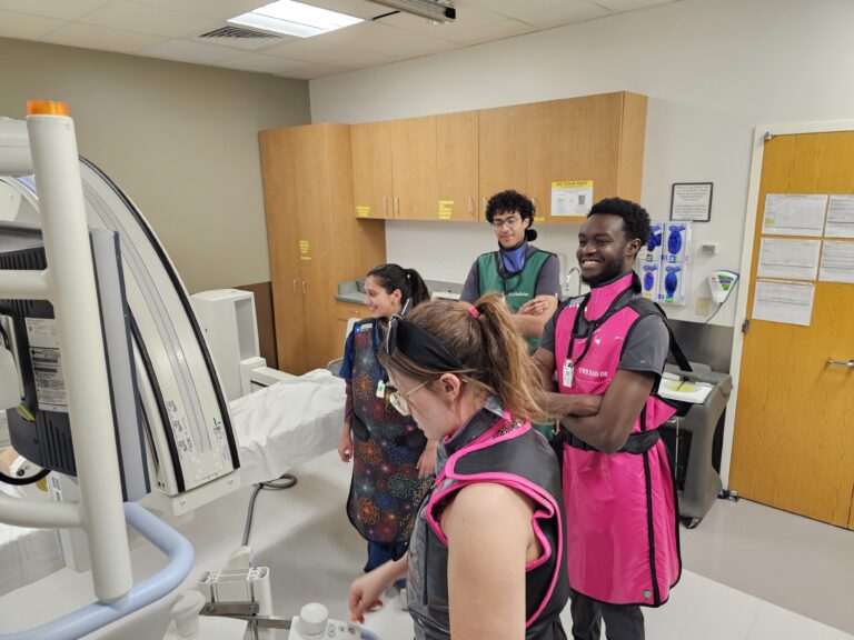
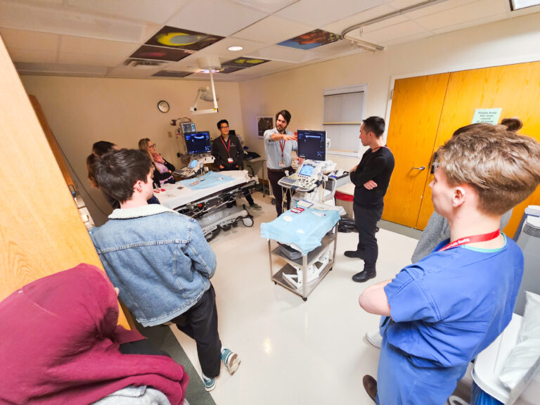
Over 30 students attended the event and were broken into three groups so they could rotate through three sessions, which were interventional radiology, breast and body ultrasound intervention, and spine imaging. Within each session, stations were set up to focus on an individual procedure. Department faculty and trainees guided students through each station, offering real-time instruction and insight into their day-to-day practices.
Interventional Radiology Session
Led by Dr. Golden, the interventional radiology session showcased complex image-guided vascular and non-vascular procedures. Students performed a mock artery embolization using an endovascular simulator at one station.
Another station demonstrated cryoablation, a minimally invasive medical procedure in which extreme cold can be used to freeze diseased tissue, such as malignant tumors. Students saw how the process works with a tank of clear gel that had several cryoprobes in place. The demonstrators activated the cryoablation system, causing the cryoprobes to cool and form small ice balls at the ends.
Additionally, students also participated in stations that demonstrated a mechanical thrombectomy, Y-90 radioembolization, embolization coils, and G-tube placements.
“We had tremendous support in making the interventional radiology session a success,” said Dr. Golden. “The event has already prompted several students to reach out regarding shadowing.”
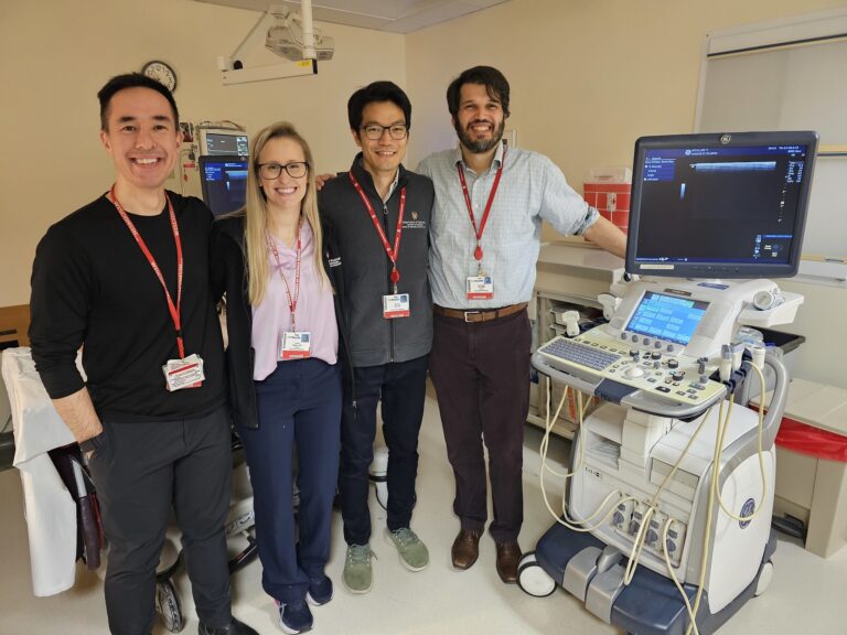
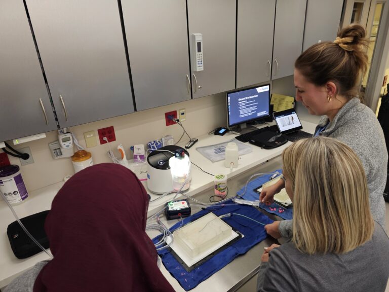
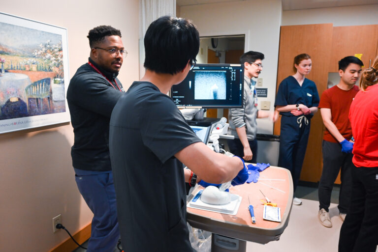
Breast and Body Ultrasound Intervention Session
This session featured demonstrations of ultrasound-guided biopsy techniques for breast, abdominal, and musculoskeletal imaging.
Thomas LoDuca, MD and breast imaging fellow Lewis Jordan, MD, MPH, MS led the breast intervention station. Dr. Jordan reflected on how this event exposed students to the practice and application of radiology in the field of medicine.
“I think seeing a program like this while in medical school actually shows how procedural the field of radiology is,” said Dr. Jordan. “It’s awesome that we are able to interpret imaging findings and use those same images to do biopsies or place localization devices. It is truly a special part of medicine.”
He hopes students walk away from this workshop with a deeper appreciation for the scope of radiology specialties.
“We’re doing more than interpreting images,” Dr. Jordan explained. “I would also hope that they would remain encouraged to learn about all of the medical specialties beyond the surface level.”
At the abdominal station for ultrasound intervention, faculty members Leslie Nelson, DO; Giuseppe Toia, MD; Matthew Lee, MD; and Michio Taya, MD demonstrated how to locate and obtain a biopsy sample by creatively using raw chicken breasts to simulate healthy tissue, with olives placed inside to represent abnormal growths or tumors. This setup helped students identify and visualize how these anomalies may appear on a scan.
Anand Narayan, MD, PhD served as a model for the musculoskeletal ultrasound demonstration. The participants witnessed how ultrasound can be used in image-guided intervention of the extremities, including arms.
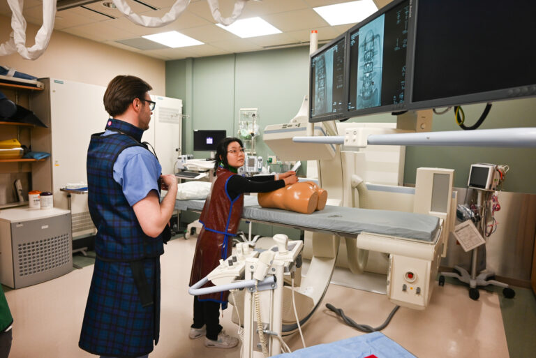
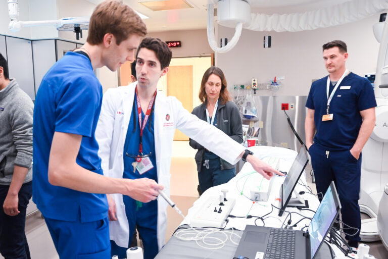
Spine Intervention Session
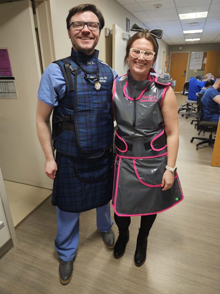
This station, led by Monica Cooley, MD and Nicholas Laucis, MD, combined diagnostic imaging with interventional pain procedures like epidural steroid injections.
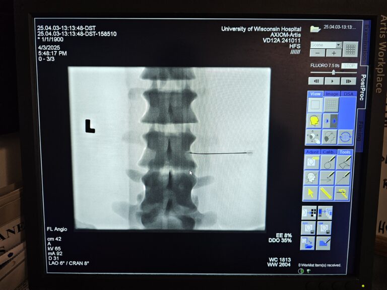
Dr. Cooley and Dr. Laucis showed students how to perform the interventional procedure using the images scanned by the C-arm as a guide for administering epidural steroid injections on a mannequin. An anatomical model of the spine was also used as a visual aid when describing and demonstrating the interventional procedure. Students administered the injection and received guidance and encouragement from their instructors and peers.
Shaping Future Radiologists Through Experience
Medical students had positive feedback about the workshop. Many noted this was their first time handling ultrasound probes or simulating image-guided procedures, and several shared that the experience sparked or deepened their interest in radiology. Faculty also appreciated teaching and interacting with students in a dynamic, low-pressure setting.
Dr. Anderson emphasized the importance of events like No Scalpel, No Problem and hands-on learning early in medical training.
“It breaks down stereotypes about the field and shows the active, procedure-oriented side of radiology,” she said. “For many students, this kind of exposure helps radiology become a more tangible career path—rather than something abstract behind a screen.”